Quantcast
Channel: Obstetrics Notes – Geeky Medics
Viewing all 57 articles
Browse latest View live

Stages of Labour

$
0
0

Childbirth is the culmination of a human pregnancy or gestation period with the birth of one or more newborn infants from a woman’s uterus.  The process of normal childbirth is categorized in three stages of labour: the shortening and dilation of the cervix, descent and birth of the infant, and birth of the placenta.

Stage 1

Onset of regular painful contractions, effacement of the cervix and dilation of the cervical Os to full dilatation.

Latent phase - cervix dilates slowly for the first 3 cm – may take hours.

Active phase- average cervical contraction is 1cm/hr in nulliparous women and 2cm/hr in multiparous women.

Stage 2

From full dilatation of the Os to the birth of the baby.

Passive stage - full dilatation → head reaches the pelvic floor and women experience a desire to push.

Active stagewhen mother is pushing, pressure of the head on the pelvic floor produces a desire to bear down and push with contractions.

Stage 3

From the birth of the baby to the expulsion of the placenta & membranes.

Takes around 15 minutes on average

Small amount of blood and the cord lengthens.

Uterine contractions compress blood vessels supporting the placenta which shears away from the wall.


Obstetric History Taking

$
0
0

It’s really important to be able to take an thorough obstetric history, because missing something could potentially have a huge impact on both mother and unborn child.  This particular history has a number of questions that are not part of the standard history taking format and therefore it’s important to be aware of just what information an obstetric history requires.

Presenting complaint

Give the patient time to explain the problem/symptoms they’ve been experiencing. 

You can then move on to more focused questioning.

Pregnancy history

Last Menstrual Period – first day
EDD – dates or scan (LMP + 9 months + 7 days)
Menstrual cycle

  • Regularity
  • Duration (normal 28 day cycle?)
  • Flow? – heavy/light – can be useful to ask number of sanitary towels/tampons

Contraceptive use? – COCP, POP, Depot, Implant
How was pregnancy confirmed?
Any other scans or tests whilst been pregnant?
– dating scan, anomaly scan

Symptoms of Pregnancy

Nausea / Vomiting - if severe known as hyperemesis gravidarum
Urinary frequency – pressure on the bladder causes this – rule out UTI
Tiredness
Breast Changes –
increase in glandular tissue – lactation
Fetal Movements - usually felt at around 18-20 weeks gestation, earlier in multips
Cravings

Ideas, Concerns & Expectations…

Previous Obstetric History

Gravidity – number of times a woman has been pregnant, regardless of outcome

Parity –  X = (any live or still birth after 24 weeks) |    Y = (number lost before 24 weeks)


Details of each pregnancy:

  • Date / Year
  • Place of birth
  • Gestation
  • Mode of delivery


Babysex, weight, current health

Problems during antenatal, labour & postnatal

Miscarriages & Terminations

Past Gynae History

Last Cervical Smear – was the result normal? – CIN?


Any gynae surgery:

- Loop excision of transitional zone (LETZ) -↑ risk of cervical incompetence
- Previous C-sections - ↑ risk of uterine rupture / placenta accreta /adhesions


Gynae investigations & treatment for:

- Infertility
- Ectopic – ↑ risk of future ectopics
- PID - chlamydia is most common cause – ↑ risk of ectopic

Past Medical History

Any hospital admissions?- when, where & why?
Any operations – abdominal or gynae?

 
Other medical conditions:

Thromboembolic disease – previous PE/DVT – high risk in following pregnancy
Diabetes – tight glycaemic control is essential – congenital defects, macrosomia 
Epilepsy – some medications are teratogenic - needs neurologist input
Hypothyroidism – TFT’s need close monitoring to keep them normal – Cretinism
Hypertension – patients BP may rise through pregnancy – PIH/Pre-eclampsia

Drug History

Pregnancy medication - folates, iron, anti-emetics, antacids

Teratogenic drugs – avoid at all costs - ACEi, Retinoids, Sodium Valproate, Methotrexate

OTC Drugs - make sure to ask patient about these, to ensure nothing unsafe

Recreational drug use – cocaine can cause placental abruption, alcohol causes FAS

ALLERGIES

Family History

Medical conditions - gestational diabetes
Inherited genetic conditions – CF
Pregnancy Loss - 
recurrent miscarriages in mother & sisters 
Pre-eclampsia - in mother or sister? – increased risk

Social History

Smoking – How many smoked and for how long? – strongly encourage to stop

AlcoholGet specific! – How much?, How often?, Everyday? Type of alcohol?

Drug use- Recreational, IV

Living SituationHouse or bungalow? Who lives with you? Is there support if needed?

Relationship Status – Married, Single

Activities of Daily Living - How are you coping at home?

Occupation - Maternity leave arranged, light duties?

Systemic Enquiry

Cardiovascular – Chest pain, PND, Orthopnoea, SOB, Cough, Ankle swelling, Palpitations, Cyanosis

Respiratory – Cough, Sputum, Haemoptysis, Chest Pain, SOB, Tachypnoea, Hoarseness, Wheezing

GI – Appetite, Diet, Nausea, Indigestion, Dysphagia, Pain,  Bowel habit, Haematemesis, Jaundice

UrinaryFrequency, Dysuria, Polyurea, Urgency, Hesitancy, Nocturia, Back pain, Incontinence

Nervous SystemVisual/other senses, Headache, Fits/Faints, LOC, Weakness, Numbness

MusculoskeletalPain in muscles, bones & joints, swelling, gait

Dermatology - Skin changes, dryness, ulcers, rashes

Obstetric Abdominal Examination

$
0
0
Introduction

Introduce yourself

Wash Hands

Ask if patient has any pain anywhere before you begin

Explanation

Describe what you are going to do

“I would like to perform an examination by gently feeling your tummy.  Although it may be a little uncomfortable, it should not be painful”
.

Gain consent

“Is that ok?”
.

Get a chaperone!!

“By law i’m required to have a female chaperone present, is that ok?”
“Do you need to empty your bladder before i examine you?”
Inspection

Position patient to the supine position

Expose abdomen from xiphisternum to the pubic symphysis

Inspect the abdomen

Asymmetry

Foetal Movements

Surgical Scars

Cutaneous Signs of Pregnancy:

  • Linea Nigra
  • Striae Gravidarum
  • Striae Albicans
Palpation

Ask about tenderness before starting!

Measure symphyseal-fundal height
  • Palpate using ulnar border of left hand moving from sternum downwards
  • Locate the fundus of the uterus (firm feeling)
  • Locate upper border of pubic symphysis
  • Measure distance in cm
  • The distance should correlate with gestational age (+/- 2cm)
Determine foetal lie

Fundal Palpation

Do this with both hands to identify which pole of foetus is at fundus


Lateral Palpation

Palpate either side of uterus, moving down to determine where the fetal back lies

.

Pelvic Manoeuvre

Turn to face patients feet

Press down on either side of lower uterus to determine presenting part

The head will be ballotable (be gentle)


How many 5th’s palpable?

Foetal head is divided into fifths in the coronal plane

If you are able to feel the entire head in the abdomen it is 5/5th’s palpable (not engaged)

If you are not able to feel the head at all abdominally it is 0/5th’s palpable (fully engaged)

Auscultation

Use a Pinard’s stethoscope

You have already identified the fetuses back

Place over anterior fetal shoulder (scapulae)

To complete the examination

Re-cover patient - allow patient to re-dress in privacy

Thank Patient

Wash Hands

Summarise Findings - “fundal heigh is 36cm which is in keeping with the current gestation.  The foetus is in a longitudinal lie, with cephalic presentation, 2/5th’s palpatable with a regular heart rate”

.

How the Gonadal Axis Works

$
0
0
What is the gonadal axis?

The gonadal axis comprises of the interaction between the hypothalamus, pituitary gland and the gonads.  The system works together to regulate development, reproductionageing and many other body processes.  It’s regulation relies upon a number of complex negative feedback loops which when lost result in disease.

How the male gonadal axis works

1. The hypothalamus secretes GnRH

2. GnRH travels down to the anterior pituitary gland
3. It binds to receptors on the pituitary gland
4. This causes release of LH (luteinizing hormone) & FSH (follicle stimulating hormone)
5. LH & FSH travel in the blood stream to the testicles
6. LH stimulates Leydig cells in the testicles to produce testosterone
  • Testosterone is required for spermatogenesis and many other important biological processes
7. FSH stimulates Sertoli cells to produce androgen binding globulin (ABG) & inhibin
  • ABG is a protein which binds to testosterone & keeps it within the seminiferous tubules
  • Inhibin helps support spermatogenesis and inhibits production of FSH, LH and GnRH

8. levels of testosterone & inhibin cause -ve feedback on the pituitary & hypothalamus


9. This results in decreased production of LH & FSH


10. As a result production of testosterone & inhibin is also decreased


How the female gonadal axis works

1. The hypothalamus secretes GnRH

2. GnRH travels down to the anterior pituitary gland

3. It binds to receptors on the pituitary gland

4. This causes release of LH (luteinizing hormone) & FSH (follicle stimulating hormone)

5. LH & FSH travel in the blood stream to the ovaries

6. When LH & FSH bind to the ovaries they stimulate production of oestrogen & inhibin

  • Oestrogen helps regulate the menstrual cycle & is essential in many body processes
  • Inhibin causes inhibition of activin which is usually responsible for stimulating GnRH production

7. Increasing levels of oestrogen & inhibin cause -ve feedback on the pituitary & hypothalamus

8. This leads to decreased production of GnRH, LH & FSH

9. This in turn results in decreased production of oestrogen and inhibin


Pre-eclampsia

$
0
0
What is Pre-eclampsia?

Pre-eclampsia is a disease of pregnancy involving the development of both hypertension as well as proteinurea. The disease appears to be caused by release of various factors from the placenta which results in widespread endothelial cell dysfunction & organ damage. Pre-eclampsia can develop anytime from 20 weeks gestation until up to 6 weeks post-partum. The earlier pre-eclampsia develops the higher the risk of serious complications. The only way to cure pre-eclampsia is to deliver the baby & remove the placenta as this is central to the disease process. Pre-eclampsia is a major cause of poor pregnancy outcome for both mother & foetus and is associated with significant mortality, therefore early recognition & intensive management can make a huge difference.

What is the mechanism of Pre-eclampsia?

The most accepted theory currently suggests the following mechanism¹,²;

1. The placenta (trophoblast) does not implant adequately into the uterine wall

2. As a result the blood supply to the placenta is relatively poor

3. This results in under-perfusion of the placenta causing hypoxia of the placental tissue

4. The hypoxic placenta releases inflammatory factors triggering an exaggerated immune response

5. The immune attack results in further release of inflammatory mediators & substances causing vasoconstriction

6. As a result the mothers blood vessels endothelial lining is damaged & peripheral vascular resistance increases

7. Increased peripheral vascular resistance results in the development of hypertension

8. Hypertension causes further damage to blood vessels & major organs including the kidney’s, which leak protein as a result

Risk factors
Patient factors

Maternal age >40

Ethnicity - african american’s have increased risk³

ObesityBMI > 35

Family history of pre-eclampsiamother or sister

Pregnancy factors

Previous pre-eclampsia

First pregnancy

Multiple pregnancy

Change of partner

Assisted reproduction techniques

Pre-existing conditions

Obesity

Diabetes mellitus

Chronic hypertension

Chronic renal disease

Thombophillia – anti-phospholipid syndrome

Symptoms & Signs
Mild

Hypertension

Significant proteinurea (>0.3g in 24hrs)

Often women are not aware of any symptoms in the early stages


Severe

Severe headache – usually frontal

Epigastric pain

Vomiting

Severe oedema - face, hands, legs, feet

Papilloedema – flashing lights, reduced vision

Reduced foetal movements - foetal distress

Intrauterine Growth Restriction

.

HELLP syndrome

HELLP syndrome can develop in severe pre-eclampsia

It is a serious disorder involving the liver & the clotting system

It’s main features are;

  • Haemolysis
  • Elevated Liver enzymes
  • Low Platelets


Diagnosis

Both Hypertension & Proteinurea must be present for the diagnosis of Pre-eclampsia to be confirmed

Hypertension

A pregnant women may classed as having hypertension in a number of ways¹

These include;

  • 140/90mmHg on 2 occasions more than 4 hours apart
  • Systolic BP of 30mmHg above the booking systolic BP
  • Diastolic BP of 15-25mmHg above booking diastolic BP


Proteinurea

>0.3g in 24-hrs

This approximates to 1+ or more on a urine dipstick

Investigations

Blood pressure monitoring

Urinalysis - protein, microscopy, culture & sensitivities

24 hour urine collection

FBC - monitor RBC for drops suggesting haemolysis

LFT – monitor liver enzymes for signs of HELLP syndrome

U&E – monitor renal function (hypertension can cause renal failure)

Clotting – if suspicious of HELLP

Management
Regular monitoring

Regular checking of;

  • BP – hypertension
  • Urine – proteinurea

.

Admission to hospital

Women may need admitting to hospital if they display any of the following¹;

  • If BP >170/110mmHg or 140/90mmHg with 2+ proteinurea
  • Significant symptoms
  • Abnormal biochemistry/ haematology
  • Significant proteinurea
  • Requires antihypertensive treatments
  • Signs of foetal compromise


Antihypertensives

The aim is to reduce the diastolic BP to <100mmHg¹

  • Labetaloloral/IV
  • Nifedipineoral
  • HydralazineIV

Avoid use of Atenolol, ACE Inhibitors or Angiotensin receptor blockers

.

Anti-seizure medication

Given if there is significant risk of eclampsia

  • Magnesium sulphate - IV – Membrane stabiliser

It halves the risk of eclampsia!¹

.

Fluid restriction

Vascular permeability is increased in pre-eclampsia

As a result fluid excess fluid spills into the extravascular compartments

If too much fluid is given it can result in pulmonary oedema

If too little fluid is given it can result in renal failure

Therefore controlling fluid intake reduces risk of fluid overload or depletion

.

Delivery of baby

Delivery is the only cure for pre-eclampsia
However the woman needs to be stable before this can be done
If foetus is <34 weeks then steroids should be given
Delivery can be delayed only if pre-eclampsia is controlled – regular monitoring required
Delaying delivery may improve perinatal outcome but increases risk to the mother
A balance must be struck between foetal & maternal wellbeing

Eclampsia

Eclampsia is a life threatening complication of pregnancy which can occur if pre-eclampsia is not controlled & becomes severe. It is characterised by the appearance of tonic clonic seizures. Eclampsia can cause coma & death in a very short period of time, therefore immediate action should be taken³.

Symptoms

Tonic Clonic Seizure:

  • Sudden loss of consciousness
  • Tonic Phase - body becomes rigid & lasts for 15-20 seconds
  • Clonic Phase - rapid contraction & relaxation of muscles causing forceful jerking of entire body

.

Coma

Death - 1 in 50 women will die from eclampsia

.

Effect’s on the foetus

Foetal distress

Bradycardia

Reduced variability on CTG

.

Placental abruption

High risk of abruption in eclampsia

The foetus becomes hypoxic as a result

Death or serious brain damage can occur

Management

ABC

Place patient on left side – avoids aortocaval compression¹

Secure airway

Give high flow oxygen

.

Antihypertensives

Labetalol, Nifedipine, Hydralazine

Given at higher doses than in routine pre-eclampsia

The aim is to stabilise BP before delivering baby via C-section

.

Magnesium sulphate

Has membrane stabilising effects reducing neuronal excitability

It halves the risk of eclampsia

Also reduces maternal mortality

.

Delivery of baby

C-section is used as it is less demanding on the body & much quicker

Carried out as soon as women is stable

Even if foetus is very premature you have to deliver

Otherwise mother will undergo multiple organ failure & die

References

1. Drife JO, Magowan (eds). Clinical Obstetrics and Gynaecology, chapter 39, pp 367-370. ISBN 0-7020-1775-2.

2. Courtney Reynolds, MD, William C. Mabie, MD, & Baha M. Sibai, MD (2006). “Preeclampsia”Pregnancy – Hypertensive Disorders. Armenian Medical Network. Retrieved 2006-11-23.

3. Douglas KA, Redman CW (November 1994). “Eclampsia in the United Kingdom”BMJ 309 (6966): 1395–400. PMC 2541348.PMID 7819845.

.

The Menstrual Cycle

$
0
0
What is the menstrual cycle?

The menstrual cycle is a complex series of physiological changes occurring in women on a monthly basis.  It results in production of an ovum & thickening of the endometrium to allow for implantation if fertilisation should occur.  The menstrual cycle is orchestrated by the endocrine system through the complex interaction of the hypothalamus, pituitary and gonads.  The entire cycle lasts around 28 days, with the cycle beginning on the first day of menstruation & ovulation occurring around day 14.


How is it controlled?


1. The hypothalamus produces Gonadotrophin Releasing Hormone (GnRH)

2. This binds to the pituitary stimulating release of;

  • Luteinizing hormone (LH)
  • Follicle Stimulating Hormone (FSH)

.

3. FSH binds to the ovaries stimulating;

  • Development of  ovarian follicles
  • Secretion of oestrogen
  • Secretion of inhibin

The follicle most sensitive to FSH becomes dominant & is known as the Graafian follicle

.

4. LH binds to the ovaries causing;

  • Production of oestrogen which is required for ovulation & thickening of the endometrium
  • Conversion of the Graafian follicle into the progesterone producing corpus luteum
  • Progesterone causes the endometrium to become receptive to implantation of a fertilised ovum

.

5. Oestrogen, Progesterone & Inhibin all cause -ve feedback on the pituitary & hypothalamus

6. This results in reduction of GnRH, FSH & LH production

7. In pregnancy GnRH, FSH & LH all remain inhibited, causing cessation of menstruation


Phases of the menstrual cycle


Follicular Phase

1. At the start of the cycle levels of FSH rise causing stimulation of a few ovarian follicles

2. As follicles mature they compete with each other for dominance

3. The 1st follicle to become fully mature will produce large amounts of oestrogen

4. This inhibits the growth of the other competing follicles

5. The 1 follicle reaching full maturity is called the Graafian follicle (oocyte develops within this)

6. The Graafian follicle continues to secrete increasing amounts of oestrogen

.

7. Oestrogen causes;

  • Endometrial thickening
  • Thinning of cervical mucous to allow easier passage of sperm

.

8. Oestrogen also initially inhibits LH production from the pituitary gland

9. However when the ovum is mature, oestrogen reaches a threshold level which conversely causes a sudden spike in LH around day 12

10. The high amounts of LH cause the membrane of the Graafian follicle to become thinner

11. Within 24-48 hours of the LH surge, the follicle ruptures releasing a secondary oocyte

12. The secondary oocyte quickly matures into an ootid & then into a mature ovum

13. The ovum is then released into the peritoneal space  & is taken into the Fallopian tube via fimbriae (finger like projections)


Luteal Phase

14. Once ovulation has occurred the hormones LH & FSH cause the remaining graffian follicle to develop into the corpus luteum
15. The corpus luteum then begins to produce the hormone progesterone

.

16. Increased levels of progesterone result in;

  • Endometrium becoming receptive to implantation of the blastocyst
  • Increased production of oestrogen by the adrenal glands
  • Negative feedback causing decreased LH & FSH (both needed to maintain the corpus luteum)
  • Increase in the woman’s basal body temperature
    .

17. As the levels of FSH & LH fall, the corpus luteum degenerates

18. This results in progesterone no longer been produced

18. The falling level of progesterone triggers menstruation & the entire cycle starts again

19. However if an ovum is fertilised it produces hCG which is similar in function to LH

20. This prevents degeneration of the corpus luteum (continued production of progesterone)

21. Continued production of progesterone prevents menstruation

22. The placenta eventually takes over the role of the corpus luteum (from 8 weeks)


The Uterine Cycle

The uterus has it’s own cycle which is driven by the cyclical release of hormones by the ovaries which we’ve previously talked about.  The inside lining of the uterus is known as the endometrium. The endometrium is the part of the uterus most affected by these changes in hormone levels.

.

It is composed of 2 layers;

  • Functional layer – this grows thicker in response to oestrogen & is shed during menstruation
  • Basal layer - this forms the foundation from which the functional layer develops – it is not shed


Phases of the uterine cycle

The uterine cycle has 3 phases known as the proliferative, secretory & menstrual phases

Proliferative phase

During the proliferative phase the endometrium is exposed to an increase in oestrogen levels caused by FSH & LH stimulating the ovaries. This oestrogen causes repair & growth of the functional endometrial layer allowing recovery from the recent menstruation & further proliferation of the endometrium.

.

Continued exposure to increasing levels of oestrogen causes;

  • Increased endometrial thickness
  • Increased vascularity -spiral arteries grow into the functional endometrial layer
  • Development of increased numbers of secretory glands

.

Secretory phase

The secretory phase begins once ovulation has occurred

This phase is driven by progesterone produced by the corpus luteum

It results in the endometrial glands beginning to secrete various substances

These secretions make the uterus a more welcoming environment for an embryo to implant

.

Menstrual phase

At the end of the luteal phase the corpus luteum degenerates (if no implantation occurs)

The loss of the corpus luteum results in decreased progesterone production

The decreasing levels of progesterone cause the spiral arteries in the functional endometrium to contract

The loss of blood supply causes the functional endometrium to become ischaemic & necrotic

As a result the functional endometrium is shed & exits out through the vagina

This is seen as the 3-5 day period of menstruation a woman experiences each month

Window of fertility

A woman’s most fertile period is between 5 days before ovulation until 1 to 2 days after

Women can therefore use knowledge of their cycle to improve chances of conception

Women may also monitor symptoms that suggest they are about to ovulate such as;

  • Basal body temperature measuringit spikes during the LH surge  24-48 hours before ovulation
  • Thinning of cervical mucous
Symptoms experienced in the menstrual cycle

Abdominal pain & cramps

Heavy vaginal bleeding

Vaginal pain

Nausea

Diarrhoea

Sweating

Fatigue

Irritability

Dysphoria (unhappiness)

References

1. Uterine cycle image - http://legacy.owensboro.kctcs.edu/gcaplan/anat2/notes/Image719.gif

How to read a CTG

$
0
0
What is Cardiotocography?

Cardiotocography (CTG) is used in pregnancy to monitor both the foetal heart as well as the contractions of the uterus. It is usually only used in the 3rd trimester. It’s purpose is to monitor foetal well-being & allow early detection of foetal distress. An abnormal CTG indicates the need for more invasive investigations & ultimately may lead to emergency caesarian section.

How it works

The device used in cardiotocography is known as a cardiotocograph.

It involves the placement of 2 transducers on the abdomen of a pregnant women.

One transducer records the foetal heart rate using ultrasound.

The other transducer monitors the contractions of the uterus.

It does this by measuring the tension of the maternal abdominal wall.

This provides an indirect indication of intrauterine pressure.

The CTG is then assessed by the midwife & obstetric medical team.

How to read a CTG

To interpret a CTG you need a structured method of assessing it’s various characteristics.

The most popular structure can be remembered using the acronym DR C BRAVADO


DRDefine Risk
CContractions
BRaBaseline Rate


VVariability

A – Accelerations

DDecelerations
O - Overall impression

Define risk

You first need to assess if this pregnancy is high or low risk

This is important as it gives more context to the CTG reading

e.g. If the pregnancy is high risk, your threshold for intervening may be lowered

Reasons a pregnancy may be considered high risk are shown below¹

.

Maternal medical illness

Gestational diabetes
Hypertension
Asthma

.

Obstetric complications

Multiple gestation
Post-date gestation
Previous cesarean section
Intrauterine growth restriction
Premature rupture of the membranes
Congenital malformations
Oxytocin induction/augmentation of labor
Pre-eclampsia

.

Other risk factors

No prenatal care
Smoking

Drug abuse

Contractions

Record the number of contractions present in a 10 minute period - e.g. 3 in 10

Each big square is equal to 1 minute, so you look how many contractions occurred in 10 squares

Individual contractions are seen as peaks on the part of the CTG monitoring uterine activity

You should assess contractions for the following:

  • Durationhow long do the contractions last?
  • Intensityhow strong are the contractions? (assessed using palpation)

.²

In this example there are 2-3 contractions in a 10 minute period - e.g. 3 in 10

..

Baseline rate of foetal heart

The baseline rate is the average heart rate of the foetus in a 10 minute window

Look at the CTG & assess what the average heart rate has been over the last 10 minutes

Ignore any Accelerations or Decelerations

A normal foetal heart rate is between 110-150 bpm¹


Foetal Tachycardia

Foetal tachycardia is defined as a baseline heart rate greater than 160 bpm

It can be caused by:¹

  • Foetal hypoxia
  • Chorioamnionitis – if maternal fever also present
  • Hyperthyroidism
  • Foetal or Maternal Anaemia
  • Foetal tachyarrhythmia


Foetal Bradycardia

Foetal bradycardia is defined as a baseline heart rate less than 120 bpm.

Mild bradycardia of between 100-120bpm is common in the following situations:

  • Post-date gestation
  • Occiput posterior or transverse presentations


Severe prolonged bradycardia (< 80 bpm for > 3 minutes) indicates severe hypoxia

Causes of prolonged severe bradycardia are:¹

  • Prolonged cord compression
  • Cord prolapse
  • Epidural & Spinal Anaesthesia
  • Maternal seizures
  • Rapid foetal descent


If the cause cannot be identified and corrected, immediate delivery is recommended

Variability

Baseline variability refers to the variation of foetal heart rate from one beat to the next

Variability occurs as a result of the interaction between the nervous system, chemoreceptors, barorecptors & cardiac responsiveness.

Therefore it is a good indicator of how healthy the foetus is at that moment in time.

This is because a healthy foetus will constantly be adapting it’s heart rate to respond to changes in it’s environment.

.

Normal variability is between 10-25 bpm³


To calculate variability you look at how much the peaks & troughs of the heart rate deviate from the baseline rate (in bpm)

.

Variability can be categorised as: 4

  • Reassuring – ≥ 5 bpm
  • Non-reassuring – < 5bpm for between 40-90 minutes
  • Abnormal – < 5bpm for >90 minutes


..

Reduced variability can be caused by: ³

  • Foetus sleeping - this should last no longer than 40 minutes – most common cause
  • Foetal acidosis (due to hypoxia) – more likely if late decelerations also present
  • Foetal tachycardia
  • Drugs – opiates, benzodiazipine’s, methyldopa, magnesium sulphate
  • Prematurity – variability is reduced at earlier gestation (<28 weeks)
  • Congenital heart abnormalities


 .

Accelerations

Accelerations are an abrupt increase in baseline heart rate of >15 bpm for >15 seconds

The presence of accelerations is reassuring

Antenatally there should be at least 2 accelerations every 15 minutes¹

Accelerations occurring alongside uterine contractions is a sign of a healthy foetus

However the absence of accelerations with an otherwise normal CTG is of uncertain significance


Decelerations are an abrupt decrease in baseline heart rate of >15 bpm for >15 seconds

There are a number of different types of decelerations, each with varying significance

.

Early deceleration

Early decelerations start when uterine contraction begins & recover when uterine contraction stops

This is due to increased foetal intracranial pressure causing increased vagal tone

It therefore quickly resolves once the uterine contraction ends & intracranial pressure reduces

This type of deceleration is therefore considered to be physiological & not pathological³


Variable deceleration

Variable decelerations are seen as a rapid fall in baseline rate with a variable recovery phase

They are variable in their duration & may not have any relationship to uterine contractions

They are most often seen during labour & in patients with reduced amniotic fluid volume

Variable decelerations are usually caused by umbilical cord compression¹

  • The umbilical vein is often occluded first causing an acceleration in response
  • Then the umbilical artery is occluded causing a subsequent rapid deceleration
  • When pressure on the cord is reduced another acceleration occurs & then the baseline rate returns
  • Accelerations before & after a variable deceleration are known as the “shoulders of deceleration”
  • There presence indicates the foetus is not yet hypoxic & is adapting to the reduced blood flow.

Variable decelerations can sometimes resolve if the mother changes position

The presence of persistent variable decelerations indicates the need for close monitoring

Variable decelerations without the shoulders is more worrying as it suggests the foetus is hypoxic


.

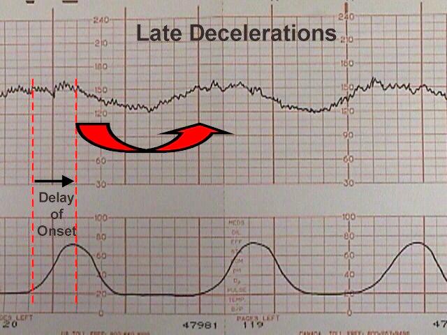
Late deceleration

Late decelerations begin at the peak of uterine contraction & recover after the contraction ends.

This type of deceleration indicates there is insufficient blood flow through the uterus & placenta

As a result blood flow to the foetus is significantly reduced causing foetal hypoxia & acidosis

.

Reduced utero-placental blood flow can be caused by: ¹

  • Maternal hypotension
  • Pre-eclampsia
  • Uterine hyper-stimulation

.

The presence of late decelerations is taken seriously & foetal blood sampling for pH is indicated

If foetal blood pH is acidotic it indicates significant foetal hypoxia & the need for emergency C-section


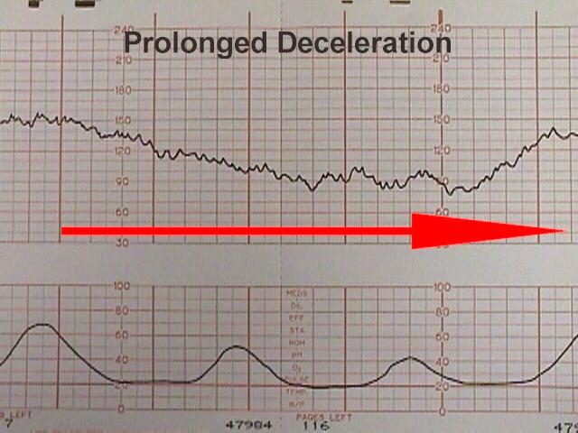
Prolonged deceleration

A deceleration that last more than 2 minutes

If it lasts between 2-3 minutes it is classed as Non-Reasurring

If it lasts longer than 3 minutes it is immediately classed as Abnormal

Action must be taken quickly – e.g. Foetal blood sampling / emergency C-section

.

Sinusoidal Pattern

This type of pattern is rare, however if present it is very serious

It is associated with high rates of foetal morbidity & mortality ¹

.

It is described as:

  • A smooth, regular, wave-like pattern
  • Frequency of around 2-5 cycles a minute
  • Stable baseline rate around 120-160 bpm
  • No beat to beat variability

A sinusoidal pattern indicates:

  • Severe foetal hypoxia
  • Severe foetal anaemia
  • Foetal/Maternal Haemorrhage

.

Immediate C-section is indicated for this kind of pattern.

Outcome is usually poor

.

Overall impression

Once you have assessed all aspects of the CTG you need to give your overall impression

The overall impression can be described as either: 4

  • Reassuring
  • Suspicious
  • Pathological

The overall impression is determined by how many of the CTG features were either reassuring, non-reassuring or abnormal. The NICE guideline below demonstrates how to decide which category a CTG falls into.4

References

Click to show

1. http://www.aafp.org/afp/990501ap/2487.html

2. http://www.fastbleep.com/medical-notes/o-g-and-paeds/16/34/449

3. Clinical obstetrics & gynaecology. 2nd Edition. 2009. B.Magowan, Philip Owen, James Drife

4. Nice guidelines http://www.nice.org.uk/nicemedia/live/11837/36273/36273.pdf


Obstetrics Quiz

$
0
0


Obstetrics Quiz

Congratulations – you have completed Obstetrics Quiz.

You scored %%SCORE%% out of %%TOTAL%%.

Your performance has been rated as %%RATING%%

Your answers are highlighted below.

Question 1
Which of the following are indications for induction of labour?
A
Risks to the mother of carrying on the pregnancy further e.g. Pre-eclampsia
B
Mum wants the baby born on her birthday at 37 weeks gestation
C
Premature rupture of membranes
D
IUGR
E
Post-term pregnancy >41 weeks
Question 2
Which of the following are drugs used to reduce the high blood pressure associated with Pre-eclampsia?
A
Lisonopril
B
Hydralazine
C
Amiodarone
D
Labetalol
E
Nifedipine
Question 2 Explanation: 
In Pre-eclampsia the aim is to control the mothers blood pressure until the pregnancy reaches a gestation at which it would be safe to deliver. However there is a fine balance between ensuring a pregnancy reaches a viable gestation & ensuring the mothers health isn’t compromised. The aim is to keep the diastolic BP <110mmHg. Labetalol, Nifedipine & Hydralazine can allow the BP to remain at a safe level for a while longer giving a few more weeks for a foetus to mature before delivery.
Question 3
At 22 weeks gestation where would you expect to find the uterine fundus?
A
Half way between umbilicus & xiphisternum
B
Symphysis Pubis
C
Umbilicus
D
Xiphisternum
Question 4
Which of the following statements are true about Magnesium Sulphate in relation to Pre-eclampsia & Eclampsia?
A
Increased tendon reflexes are an early sign of Magnesium Sulphate overdose
B
IV Magnesium Sulphate is indicated if there is significant risk of a mother developing Eclampsia.
C
Magnesium Sulphate acts a membrane stabiliser, reducing the excitability of neurones.
D
Magnesium Sulphate halves the risk of Eclampsia
E
Decreased tendon reflexes are an early sign of Magnesium Sulphate overdose
Question 4 Explanation: 
Eclampsia is a life threatening complication of pregnancy which can occur if pre-eclampsia is not controlled & becomes severe. It is characterised by the appearance of tonic clonic seizures. Eclampsia can cause coma & death in a very short period of time, therefore immediate action should be taken. Magnesium Sulphate is given to those at significant risk of developing Eclampsia.
Question 5
Which of the following symptoms are experienced by women suffering with severe pre-eclampsia?
A
Epigastric pain
B
Increased appetite
C
Vomiting
D
Swollen face, hands & feet
E
Frontal Headache
Question 5 Explanation: 
In severe pre-eclampsia the symptoms are widespread & affect many different body systems. The high blood pressure leads to increased intracranial pressure causing headache & papilloedema which causes visual disturbances. If a women presents with these symptoms immediate action must be taken as their is a significant risk of death for both the mother and foetus.
Question 6
When screening for Down’s syndrome which 3 of the following does the “Combined Test” involve?
A
Measurement of PAPP-A
B
Measurement of Nuchal Translucency
C
Measurement of Alpha Fetoprotein (AFP)
D
Measurement of Unconjugated Estriol (uE3)
E
Measurement of Free Beta hCG
Question 6 Explanation: 
All women presenting before 20 weeks of pregnancy should be offered screening tests for Down’s syndrome regardless of their age. The “Combined Test” is usually carried out between 11 weeks to 13 weeks + 6 days gestation.

If the women has not presented to late to have the combined test they can have “Serum Screening” carried out between 15-20 weeks. There are a number of different types of “Serum Screening” such as Double, Triple & Quadruple tests. The Quadruple test measures free Beta hCG, AFP, Inhibin-A & uE3.

Question 7
What does asymmetrical Intra-uterine Growth Restriction (Normal head circumference with reduced abdominal circumference) suggest?
A
Chromosomal disorder
B
Placental insufficiency
Question 7 Explanation: 
If the placenta is not supplying adequate blood to the foetus there will be reduced amounts of energy for the baby to use. As a result the body directs the remaining fuel to allow normal brain development at the expense of the abdomen (glycogen stores in the liver) As a result the abdominal circumference decreases however the head circumference remains normal.
Question 8
Which of the following are risk factors for Post Partum Haemorrhage?
A
Multiple pregnancy
B
Pre-eclampsia
C
Asian ethnicity
D
Nulliparity
E
Antepartum haemorrhage in current pregnancy
F
Previous menorrhagia
G
Placenta praevia
H
Previous endometriosis
Question 8 Explanation: 
There are many significant risk factors for PPH. The amount each increases risk is as follows;
- Placenta praevia (15x)
- Multiple pregnancy (5x)
- Pre-eclampsia (4x)
- Nulliparity (3x)
- Previous PPH (3x)
- Asian Ethnicity (2x)
- Obesity (2x)
Question 9
The “Serum Screening” test for Down’s syndrome is carried out between 15-20 weeks for those women who presented too late to have the “Combined Test”.  In the “Quadruple Serum Screening Test” which 4 of the following are measured?
A
Unconjugated Estriol (uE3)
B
Inhibin-A
C
Serum insulin
D
Alpha Fetoprotein (AFP)
E
Free Beta hCG
Question 10
What is a mothers risk of having a child with Down’s syndrome at the age of 20?
A
1 in 270
B
1 in 50
C
1 in 100
D
1 in 1500
E
1 in 800
Question 10 Explanation: 
Down’s syndrome is rarely seen in younger mothers & as a result many opt to not have screening for Down’s syndrome during pregnancy.
Question 11
What period of gestation does the 2nd trimester represent?
A
10-20
B
15-30
C
14-29
D
13-28
Question 11 Explanation: 
The second trimester begins at 13 weeks and ends at 28 weeks.
Question 12
What period of gestation does the 3rd trimester represent?
A
28-40 weeks
B
27-39 weeks
C
30-41 weeks
D
29-40 weeks
Question 12 Explanation: 
The third trimester starts at 29 weeks & ends at 40 weeks.
Question 13
Which of the following is a prostaglandin commonly used in induction of labour
A
Atenolol
B
Labetalol
C
Misoprostol
Question 13 Explanation: 
Misoprostol is inserted vaginally & causes effacement of the cervix & uterine contractions.
Question 14
At 36 weeks gestation where would you expect to find the uterine fundus?
A
Xiphisternum
B
Symphysis Pubis
C
Halfway between Umbilicus & Xiphisternum
D
Umbilicus
Question 15
Women with mild pre-eclampsia usually present to their doctor due to significant symptoms
A
False
B
True
Question 15 Explanation: 
Women with mild pre-eclampsia often do not have any symptoms at all. As a result they are usually picked up as a result of blood pressure screening by their midwife.
Question 16
What does symmetrical Intra-uterine Growth Restriction (both the head circumference & abdominal circumference are lower than normal) suggest?
A
Placental insufficiency
B
Chromosomal Disorder
Question 16 Explanation: 
Symmetrical IUGR usually suggests a baby has a chromosomal disorder. This is because in a healthy baby the head circumference should remain normal, even if there is significant placental insufficiency, as the body makes sure the brain receives the necessary fuel at the expense of the rest of the body. That’s why in placental insufficiency the abdominal circumference is reduced but the head circumference is normal (asymmetrical IUGR)
Question 17
At what stage of gestation would you expect a nulliparous women to begin to feel foetal movements?
A
18-20 weeks
B
14-16 weeks
C
10-12 weeks
D
22-24 weeks
Question 17 Explanation: 
Most women will become aware of foetal movements around 18-20 weeks. However if a women has had previous pregnancies they tend to notice movements earlier around 15-18 weeks.
Question 18
Which of the following statements about Pre-eclampsia are correct?
A
If Pre-eclampsia is treated early & aggressively with antihypertensives it can be cured.
B
The main driver of pathological process of Pre-eclampsia is the interaction between the immune system & placenta.
C
The only way to cure Pre-eclampsia is by delivering the baby & removing the placenta.
Question 19
Which of the following methods are the correct way to calculate the Estimated Date of Delivery (EDD)?
A
First day of LMP + 8 months & 1 week
B
First day of LMP + 9 months
C
Last day of LMP + 8 months & 1 week
D
First day of LMP + 9 months & 1 week
Question 19 Explanation: 
The correct way to calculate the EDD is to add 9 months & 1 week onto the first day of the last normal menstrual period. This method may not be reliable if a women is unsure about when her last menstrual period was. As a result ultrasound is now used to date pregnancies.
Question 20
Which of the following are causes of Post Partum Haemorrhage?
A
Vaginal or Vulval lacerations
B
Uterine Atony
C
Retained Placenta
D
Coagulapathy
Question 20 Explanation: 
All of the above are causes of PPH with uterine atony been the most common. Coagulapathy accounts for only around 1% of PPH however it’s important to keep it in mind so you don’t miss it.
Question 21
At 12 weeks gestation where would you expect to feel the uterine fundus?
A
Xiphisternum
B
Umbilicus
C
Half way between umbilicus & xiphisternum
D
Symphysis pubis
Question 22
Which 3 of the following statements about Chorionic Villus Sampling are correct?
A
The risk of pregnancy loss is around 2% in the first trimester & 3% in the second trimester
B
Chorionic Villus Sampling involves sampling part of the developing placenta
C
Chorionic villus sampling is usually performed between 11 to 13 weeks + 6 days.
D
Chorionic villus sampling is usually performed between 13 to 15 weeks + 6 days.
E
Chorionic Villus Sampling involves sampling the amniotic fluid which surrounds the foetus
Question 22 Explanation: 
Chorionic Villus Sampling involves the sampling of developing placental tissue. It is the procedure of choice at earlier gestations (before 12 weeks) as it carries a lower risk of pregnancy loss compared to Amniocentesis. However in the second trimester Amniocentesis is favoured as it carries a lower risk of pregnancy loss.
Question 23
Which of the following is thought to be a cause of Hyperemesis Gravidarum?
A
Underlying infection
B
Psychological issues
C
High levels of circulating HCG
D
Over eating
Question 23 Explanation: 
Hyperemesis Gravidarum is a severe form of morning sickness in which women suffer with excessive nausea & vomiting which prevents them taking in adequate amounts of food & water. It is thought to be caused by high levels of circulating HCG. This is supported by the fact that it’s more common in multiple pregnancies & also tends to only affect women during the first 12 weeks of pregnancy.
Question 24
Which 2 of the following does a membrane sweep” involve?
A
Rupture of membranes
B
Use of finger to separate the amniotic membrane from the cervix.
C
Release of prostaglandins
D
Significant bleeding
Question 24 Explanation: 
A midwife usually performs an internal examination on the mother & uses their finger to separate the amniotic membranes from the walls of the cervix. This causes release of prostaglandins which can kickstart the labour process.
Question 25
Which of the following are risk factors for Pre-eclampsia?
A
Change of partner
B
Maternal age > 40
C
First pregnancy
D
Obesity (BMI >35)
E
Family history of pre-eclampsia in mother or sisters
Question 25 Explanation: 
The risk of Pre-eclampsia is influenced by many different factors. One which isn’t immediately obvious is the increased risk after changing partner. This is thought to be due to the fact that the mothers immune system is less tolerant of the new antigens from the new partner.
Question 26
What is a mothers risk of having a child with Down’s syndrome at the age of 40?
A
1 in 270
B
1 in 800
C
1 in 50
D
1 in 1500
E
1 in 100
Question 26 Explanation: 
The risk of having a child with Down’s syndrome increases with age. It’s important to know the chances a mother will have a child with Down’s syndrome so you can communicate this to your patient allowing them to make a more informed decision about pregnancy.
Question 27
What is a mothers risk of having a child with Down’s syndrome at age 46?
A
> 1 in 50
B
1 in 1500
C
1 in 800
D
1 in 270
E
1 in 100
Question 28
Which 3 of the following statements are true about Amniocentesis?
A
Amniocentesis can be carried out from 8 weeks gestation
B
Early amniocentesis (12 weeks gestation) carries a greater risk of pregnancy loss compared to Chorionic Villus Sampling at the same gestation.
C
Amniocentesis can be carried out from 12 weeks gestation
D
There is a 3% probability of losing a pregnancy as a result of Amniocentesis.
E
There is a 0.5-1% probability of losing a pregnancy as a result of Amniocentesis
Question 28 Explanation: 
Amniocentesis involves sampling the amniotic fluid around the foetus during pregnancy. It can be carried out from 12 weeks gestation (early amniocentesis) however it can also be carried out later (16-18 weeks gestation) which reduces the risk of pregnancy loss. Chorionic Villus Sampling carries a lower risk of pregnancy loss compared to early amniocentesis, however a greater risk when compared to later amniocentesis. As a result Chorionic Villus Sampling is preferred at earlier gestations & is the investigation of choice before 12 weeks gestation.
Question 29
Which of the following are complications of induction?
A
Caesarian section
B
Uterine hyper-stimulation
C
IUGR
D
Uterine rupture
E
Prolapsed cord
Question 29 Explanation: 
Induction of labour increases the risk of a number of complications which are seen in vaginal delivery. The uterus can become over stimulated causing prolonged contractions which can starve the baby of oxygen. It can also cause contractions to be so powerful they cause rupture of the uterus. Finally if a women is induced to early it can result in prolonged labour & eventually emergency C-section or an instrumental delivery.
Question 30
In severe Pre-eclampsia women can develop HELLP syndrome which is very serious & life threatening.  Which of the following are characteristic findings in the HELLP syndrome?
A
Low platelets
B
Elevated Liver enzymes
C
Elevated Potassium
D
Haemolysis
E
Elevated Sodium
Question 30 Explanation: 
HELLP syndrome is a serious complication of severe pre-eclampsia & involves derangement of liver function & the clotting system. If not treated quickly it can result in massive haemorrhage as well as liver & renal failure.
Question 31
Which of the following statements describe the first stage of labour correctly?
A
Starts when the effaced cervix is 3cm dilated and ends when the cervix is fully dilated at 10cm.
B
Starts when regular painful contractions begin & ends when the cervix is fully effaced and dilated to 5 cm.
C
Onset of painful contractions to full effacement of the cervix. The membranes are still intact
Question 32


Which sign is present on this pregnant abdomen?

A
Stria Albicans
B
Striae Gravidarum
C
Linea Nigra
Question 32 Explanation: 
This images shows a linea nigra. This occurs in around 3/4 of pregnancies. It’s described as a vertical line of increased pigmentation running down the centre of the pregnant abdomen. The increased pigmentation occurs over the linea alba which is stretched during pregnancy to accommodate the developing foetus.
Question 33
At 28 weeks gestation where would you expect to feel the uterine fundus?
A
Umbilicus
B
Xiphisternum
C
Halfway between Xiphisternum & Umbilicus
D
Symphysis Pubis
Question 34
What is the most common cause of Post Partum Haemorrhage?
A
Uterine atony
B
Vulval or Vaginal Lacerations
C
Uterine rupture
D
Retained placenta
Question 34 Explanation: 
The most common cause of PPH is Uterine Atony however retained placenta & vaginal lacerations also account for a significant number of instances of PPH. Uterine atony described a state in which the uterus can no longer effectively contract. Because the uterus cannot contract all the blood vessels which were supplying the placenta are not clamped off & therefore can bleed profusely. A uterus can become atonic for a number of reasons such as prolonged labour, large baby, multiple pregnancy & retained placenta. If a women is found experiencing PPH its important to apply firm pressure to uterus immediately to reduce blood loss.
Question 35
What period of gestation does the 1st trimester represent?
A
1-11 weeks
B
1-10 weeks
C
1-12 weeks
D
1-13 weeks
Question 35 Explanation: 
The first trimester refers to the first 12 weeks of pregnancy.
Question 36
Which of the following statements describes the 2nd stage of labour correctly?
A
The second stage begins at full dilatation of the cervix and ends when the baby is born.
B
The second stage begins at full dilation of the cervix and ends once the baby & placenta have been expelled from uterus.
C
The second stage begins when the cervix are 7 cm dilated & ends when the cervix reaches full dilatation.
Question 37
Which of the following need to be present for a women to be diagnosed as having Pre-eclampsia?
A
Proteinurea
B
Hypertension
C
Haematuria
D
Visual Disturbances
E
Hypotension
Question 37 Explanation: 
Pre-eclampsia is defined as the presence of both hypertension & proteinurea during pregnancy. Hypertension alone would be considered Pregnancy Induced Hypertension.
Question 38
What is the definition of Post Partum Haemorrhage?
A
Loss of >200ml of blood from the genital tract within 24 hours of delivery
B
Loss of >500ml of blood from the genital tract within 24 hours of delivery
C
Loss of >500ml of blood from the genital tract during delivery
D
Loss of >200ml of blood from the genital tract during delivery
Question 38 Explanation: 
Primary postpartum haemorrhage is defined as a loss of blood >500ml from the genital tract within 24 hours of delivery. If the blood loss is between 500-1000ml it is defined as a Minor PPH. If the blood loss is more than 1000ml it is defined as a Major PPH & can result in death if not managed quickly & effectively.
Question 39
Which 2 of the following are drugs which can be used to induce labour?
A
Misoprostol
B
Syntocinon
C
Labetalol
D
Nitrogen Oxide
Question 39 Explanation: 
Syntocinon is a synthetic form of Oxytocin. Oxytocin causes uterine contractions & cervical dilatation. Misoprostol is a prostaglandin which causes thinning of the cervix and uterine contraction. Usually women are given a prostaglandin first to induce labour, however if prostaglandins fail they can then be given Oxytocin. However you cannot give Oxytocin if you have had any prostaglandins within 6 hours previous.
Question 40
Which of the following statements describes the 3rd stage of labour correctly?
A
The third stage begins once the baby is born & ends 24 hours after the time the baby was born.
B
The third stage begins once the baby is born and ends once the placenta and membranes have been expelled from the uterus.
C
The third stage begins once the baby & placenta have been expelled from the uterus & ends when the uterus contracts down to it’s normal size.
Question 41
If a baby is found to have Intra-uterine Growth Restriction, what might be the next steps of management?
A
Termination of pregnancy
B
Serial growth scans
C
USS scan to look for structural problems which may suggest chromosomal abnormality.
D
Umbilical artery doppler
E
Delivery if >37 weeks
Question 41 Explanation: 
Umbilical artery doppler would be a logical next step to look at the blood flow of the placenta. If there was very poor blood flow, or reverse end diastolic flow it may suggest the need for prompt delivery. However if blood flow is normal an USS may be carried out to rule of chromosomal causes of IUGR such as Down’s syndrome. The women would also require regular follow up & serial growth scans to allow the growth trends to be monitored closely for signs of deterioration. If a women is already >37 weeks delivery may be suggested as it may be more dangerous to wait a few weeks more.
Question 42
Which of the following are causes of Intra-Uterine Growth Restriction (IUGR)?
A
Pre-eclampsia
B
Diabetes
C
Alcohol
D
Hypercholesterolaemia
E
Hypertension
F
Smoking
Question 42 Explanation: 
IUGR can be caused by many different factors. One very important one is smoking which accounts for around 30-40% of cases of IUGR. Alcohol abuse also causes IUGR and foetal alcohol syndrome. It’s essential to communicate to mothers the risks of smoking & alcohol in pregnancy to allow them to make a more informed decision about their lifestyle choices during pregnancy.
Once you are finished, click the button below. Any items you have not completed will be marked incorrect.
Get Results


There are 42 questions to complete.


Postpartum Haemorrhage

$
0
0

Primary Postpartum Haemorrhage

Primary postpartum haemorrhage (PPH) is defined as loss of more than 500ml of blood from the genital tract within 24 hours of delivery¹


PPH can be described as minor or major depending upon the level of blood loss;

  • Minor PPH involves the loss of of anything up to 1000mls of blood
  • Major PPH involves the loss of more than 1000mls of blood
Causes

Causes of postpartum haemorrhage include uterine atony, trauma, retained placenta, and coagulopathy. These causes are often referred to as the ”four Ts” Tone (uterine atony) Trauma (lacerations) Tissue (retained placenta)  Thrombin (coagulopathy)2

Uterine atony

Uterine atony is the most common cause of PPH account for around 70% of cases.² It occurs due to the uterus losing its muscle tone.  As a result it does not contract after delivery and therefore the uterine vessels are not clamped down on allowing large amounts of bleeding to occur.  The uterus can become atonic for a number of reasons such as prolonged labour (the uterus tires out) or multiple birth (the uterus is large & stretched making it less effective at contracting)

Retained placenta

Retained placenta accounts for around 10% of cases of PPH.² It results from either all or part of the placenta been retained within the uterus. This prevents the uterus from contracting fully and therefore the uterine vessels are not occluded & continue to bleed.

Vaginal or Vulval Lacerations

Vulval & vaginal lacerations account for most of the other 20% of PPH cases.² They occur due to the mechanics of birth and can be very severe.  They must be monitored for some time afterwards to ensure no infection develops and proper healing occurs.

Coagulopathy

Around 1% of PPH cases occur as a result of coagulapathy.² Common causes of coagulopathy include haemophilia A or B.   It’s important to rule out coagulopathy as it can result in the death of a patient if not identified early.

Risk factors
Pregnancy factors³

Placenta praevia (15 x risk)

Multiple pregnancy (5 x risk)

Pre-eclampsia (4 x risk)

Previous PPH (3 x risk)

Nulliparity (first childbirth) (3 x risk)

Placental abruption


Delivery factors³

Emergency caesarian section (9 x risk)

Retained placenta (5 x risk)

Elective caesarian section (4 x risk)

Mediolateral Episiotomy

Prolonged labour (>12 hours)

Big baby (>4kg)


Maternal factors³

Obesity – BMI >35 (2 x risk)

Asian ethnicity

Haemophilia (type A or B)

Anaemia (<9g/dl)

Age > 40 years


Symptoms & Signs
Symptoms³

Prolonged & worsening vaginal bleeding after delivery

Passing many large clots

Lower abdominal pain

Pelvic pain

Light headedness / Fainting

Shortness of breath


Signs

Pale skin

Tachycardia

Tachypnea

Hypotension

Tender abdomen

Fever

Decreased consciousness


Investigations³

Vaginal examination – determine source of bleeding

Abdominal examination – tender abdomen suggests uterine bleeding

FBCsudden drop in Hb suggests acute blood loss

Clottingprolonged PT & APTT suggest disseminated intravascular coagulation

Blood pressurelow BP indicates blood loss

Pulse rate – usually tachycardia present due to hypovolaemic shock

Oxygen saturation – may be decreased to decreased red blood cells & Hb

Hourly urine output - if decreased suggests blood volume loss

ECG

Complications

Hypovolaemic Shock 

HELLP – Haemolysis, Elevated Liver Enzymes, Low Platelets

Disseminated Intravascular Coagulation – due to consumption of clotting factors

Management ³
Minor PPH

If minor PPH with no clinical signs of shock;

  • Contact obstetric doctor
  • Get IV access with two 14 gauge cannula’s
  • Crystalloid infusion
  • Crossmatch 2 units of blood
  • BP, Pulse & Oxygen Saturation
  • FBC
  • Clotting
  • Regular clinical monitoring & observation


Major PPH

Contact senior clinical staff

Call Obstetric registrar & alert Consultant

Call Anaesthetic registrar & alert Consultant

Experienced Midwife

Alert consultant haematologist on call

Alert blood transfusion lab

Call porters to transfer specimens & blood products quickly


Airways & Breathing

Check airway - if compromised get anaesthetist to secure airway

Give Oxygen at 10/15 L/min

.

Circulation

Secure IV access with two 14 gauge cannula’s

Blood sample should be taken & sent for;

  • FBC
  • Coagulation
  • U&E
  • Crossmatch – 4 units
  • Renal & Liver function tests


Resuscitation

Position patient in supine position

Keep patient warm - bear-hugger

Transfuse blood as soon as possiblegive 4 units of fresh frozen plasma for every 6 units of blood

.

Until blood is available infuse;

  • up to 3.5 litres of warmed crystalloid Hartmann’s solution
  • and/or 1-2 litres of colloid as rapidly as required

.

Give platelets if PLT count <50 x 109

Give cryoprecipitate if fibrinogen <1g/L


Stopping the bleeding

In uterine atony try each of the following measures in turn until bleeding stops;

  • Bimanual uterine compression (rubbing up the fundus) – stimulates contractions
  • Syntocinon - 5 units – slow IV injection
  • Ergometrine0.5mg – slow IV injection
  • Syntocinon infusion40 units in 500 ml Hartmann’s solution at 125 ml/hour
  • Carboprost - 0.25 mg by intramuscular injection every 15 mins (max of 8 doses) 
  • Carboprost - intramyometrial – 0.5mg
  • Misoprostol - 1000mg rectally


If pharmacological measures fail, surgical treatment should be initiated in the following order;

  • Intrauterine balloon tamponade – blood inside uterus to compress vessels
  • Haemostatic brace suturing - large sutures around uterus to compress it
  • Bilateral ligation of uterine arteries
  • Bilateral ligation of internal iliac arteries
  • If bleeding will not stop hysterectomy must be performed - do not hesitate with decision


Intensive monitoring

Patient should be sent to HDU or ICU

Prevention

Active management of the 3rd stage of labour reduces maternal blood loss & chances of PPH

Prophylactic oxytocics should be offered to all women as they reduce chances of PPH by 60%

  • Low risk vaginal delivery – oxytocin – 5 or 10 IU’s – IM injection
  • Caesarian section – oxytocin – 5 IU – slow intravenous infusion


 Secondary Postpartum Haemorrhage

Secondary PPH presents as abnormal bleeding from the genetical tract anytime from 24 hours after delivery until up to 6 weeks postpartum.


Causes

Infection (Endometritis) – 1-3% of spontaneous vaginal deliveries

Retained products of conception 


Examination

Tachycardia

Fever

Rigors

Tender abdomen (suprapubic)

Fundus may be elevated in cases of retained products


Investigations

FBC - low Hb may suggest significant blood loss

MSU - could give pain & haematuria which may be confused with PPH

High vaginal swab – can confirm presence of infection

USS -often can be normal despite retained products been present

Blood cultures – may show septicaemia


Management³

Speculum examination

Look for signs of inflammation

Rule out lacerations

Identify clots preventing closure of cervical OS

Clots can then be removed at the time


Antibiotics

If antibiotics are indicated combination of ampicillin and metronidazole is appropriate. 

If patient has endomyometritis (tender uterus) or sepsis, gentamicin should also be used


Surgery

Surgical measures should be undertaken if there is excessive or continuing bleeding

  • Regardless of ultrasound findings
  • Surgical evacuation of retained products of conception – consultant required


References

Click to show

1. Prevention and management of postpartum haemorrhage, Royal College of Obstetricians and Gynaecologists (May 2009)

2. Four T’s of PPH - Anderson JM, Etches D (March 2007). “Prevention and management of postpartum hemorrhage”. American Family Physician 75 (6): 875–82.

3. Postpartum Haemorrhage, Prevention and Management. Royal College of Gynaecologists. May 2009.

Physiological Aspects of Normal Pregnancy

$
0
0
Definitions for Dating Pregnancy

Conception: 2 weeks after 1st day of last menstrual period (LMP) with a regular 28 day cycle

Estimated Due Date (EDD): Naegele’s rule – add 9 months + 7 days to 1st day of LMP. Pregnancy lasts 38wks from conception/40wks from 1st day of LMP

Trimesters:

  • 1st trimester = 1-13wks
  • 2nd trimester = 14-27wks
  • 3rd trimester = 28-40wks


Puerperium:

  • Delivery –> 6wks.
  • Reversal of the physiological changes of pregnancy
Maternal Physiological Changes During Pregnancy
Cardiovascular and Respiratory Changes
  • 40% increase in plasma volume by 32wks. [RBC] increases by 20%.
  • 40% rise in cardiac output. CO and BP fall if supine due to vena cava compression.
  • Reduced peripheral venous return causes BP drop in early pregnancy. Return to pre-pregnancy level later.
  • 40% increase in tidal volume.
  • Haemodilution: overall amount of Hb rises, but concentration falls.
  • O2 demand increases by 15%.
  • Increased clotting risk: increased factor VII, VIII, X, and rise in fibrinogen.
  • Increased RBC mass: protects against the ~0.5L delivery blood loss (1L if twins or CS)
Renal changes
  • Renal pelvis + ureters dilate (pressure/progesterone): risk of acute pylonephritis.
  • GFR increases by 50%: reduces plasma urea, creatinine and osmolality.
  • Increased urinary protein loss. >500mg in 24hrs is abnormal however.
Endocrine changes
  • Insulin secretion doubles. Physiological glycosuria may occur.
  • Thyroid binding globulin doubles. T3 + T4 fall slightly. Goitre more common.
  • Anterior pituitary doubles in size – risk of ischaemia in PPH (Sheehans syndrome)
  • Rise in total and free serum cortisol and urinary free cortisol
Musculoskeletal + skin changes
  • Joints of the lower back and pelvis soften.
  • Increased incidence of rashes/epistaxis/hyperpigmentation/spider naevi/erythema
Calcium and phosphate
  •  Increased demand of Ca (especially in 3rd trimester and puerperium) leads to increased gut absorption. Calcium is actively transported across placenta.
  • Serum Ca + phosphate levels fall (bound to albumin). Ionised Ca remains stable
Liver
  • Hepatic blood flow unchanged
  • ALP levels rise by 50% and albumin falls by 10g/L (causes a fall in total protein)
Uterine physiology
  • Morula becomes blastocyst at the 32 cell stage
  • Implantation 7-14 days post conception: blastocyst attaches and trophoblast cells invade the endometrium.
  • Organogenesis: 2-8 wks post conception.
  • Inner cell mass becomes embryo. Trophectoderm becomes placental trophoblast.
  • Foetus develops in amniotic cavity, attached to placenta by umbilical cord.
  • Amnion: membrane lining of cavity, expands as placenta progresses. 2nd layer (chorion) in apposition to the amnion.
  • Placenta is anchored to maternal decidua.
  • Intervillous space supplied by maternal spiral arteries.
  • Cord has 2 arteries (deoxygenated blood from fetus to placenta) + 1 vein (oxygenated blood from placenta to foetus).
  • Uterus holds 5L at term (500x pre-pregnancy): muscle hypertrophy.
  • Blood supply from uterine + ovarian arteries.
  • Cervical mucous plug protects during pregnancy.
Multiple Pregnancy
  • 10X higher risk of stillbirth
  • 50% pre-term
  • Increased risk of IUGR

Dizygotic = non-identical, duplication of normal processes, dichorionic, diamniotic.

Monozygotic = earlier split, more independent. DCDA <3 days, 4-7days MCDA, >8d MCMA. Requires tertiary centre care if monozygotic!

Risks to mother:

  • Anaemia
  • Pre-eclampsia
  • Hyperemesis
  • Polyhydramnios
  • Complicated labour


Risks to developing foetuses:

  • Congenital abnormalities
  • Twin-to-twin transfusion
  • Pre-term delivery
  • Twin entrapment

More regular antenatal checks are required.

Foetal Growth

Growth governed by intrinsic (maternal height/weight/ethnicity, fetal sex/genes/conditions) and extrinsic (environmental – social class, nutrition, maternal disease) factors.

Small for Gestational Age – a foetus that has failed to achieve a specific biometric or estimated weight threshold by a specific gestational age – may be constitutional or due to intra-uterine growth restriction (IUGR).

Assessments of foetal growth: Biparietal distance, head circumference, abdominal circumference, femur length – serial measurements more useful to gauge velocity. Plotted on centiles – can show if any dropping off/lag/acceleration.

Assessment of baby’s coping: kick charts, CTG, biophysical profile (fetal breathing movements, fetal movements, fetal tone, amniotic fluid volume).

Assessment of baby’s nutrition: placental assessment – appearance, blood flow characteristics via Doppler (umbilical – fetus to placenta, uterine – mother to placenta). Progressively greater resistance leads to absent or reversed End Diastolic Flow.  (read up on this if you’re interested, I won’t go into it further!)

Foetal Development
Early Pregnancy
  • Day 14 = ovulation
  • Fertilisation occurs commonly in the fallopian tube
  • Cell division occurs: zygote → morula → blastocyst as moving to uterine cavity
  • Day 23 = implantation – beginning of fetal-maternal dialogue
  • When the blastocyst implants – production of hCG by the decidua stimulates the ovary to produce progesterone (causes modification of maternal physiology).
  • hCG levels rapidly rise <10wks. Can be detected in serum/urine 4 weeks after LMP (urine PT +ve when concentration of hCG is 25IU/ml)
Ultrasound
  • 4-5 weeks – gestation sac ~6mm
  • 5-6 weeks – yolk sac
  • 6 weeks – foetal pole ~5mm
  • 7 weeks – foetal heart activity
  • 8 weeks – limb buds, fetal movements
  • Foetus should double in size every week until 12 weeks gestation

The post Physiological Aspects of Normal Pregnancy appeared first on Geeky Medics.

Gynaecology Quiz

$
0
0

Put your knowledge of gynaecology to the test with this awesome fact packed gynaecology quiz!

Female Reproductive Pathology

Congratulations - you have completed Female Reproductive Pathology.

You scored %%SCORE%% out of %%TOTAL%%.

Your performance has been rated as %%RATING%%


Your answers are highlighted below.
Question 1
A unilateral and painful lesion is found in the lower vestibule, adjacent to the vaginal canal. What disease is this a description of?
A
Bartholin cyst
B
Condyloma
C
Lichen sclerosis
D
Vulvar carcinoma
Question 1 Explanation: 
A bartholin cyst is a swelling of the bartholin gland due to obstruction. A collection of pus may then form creating a painful cystic lesion as there is a more increased risk of infection.
Question 2
What is the classic finding of HPV infected cells?
A
Rhabdomyoblast
B
Koilocytes
C
Schiller Duval bodies
D
Granulomas
Question 2 Explanation: 
Koilocytes are a sign of HPV infection. They have ‘raisinoid’ nuclei, some with a perinuclear halo.
Question 3
Leukoplakia with paper thin vulvar skin, seen most commonly in post menopausal women. What disease is this?
A
Condyloma
B
Lichen simplex chronicus
C
Extramammary Paget disease
D
Lichen sclerosis
Question 3 Explanation: 
Lichen sclerosis is a condition in which there is thinning of the epidermis. It carries a small risk of developing vulvar carcinoma.
Question 4
The vulva can be affected with extra-mammary Paget disease. What is the stain for Paget cells?
A
PAS+, keratin +, S100+
B
PAS -, keratin -, S100-
C
PAS+, keratin+, S100-
D
PAS-, keratin-, S100+
Question 4 Explanation: 
It is important to differentiate between Paget’s disease and melanoma as both present with similar features on the vulva. Thus, knowledge of the stains is crucial. Choice C describes Paget’s disease whilst Choice D describes the stain for melanoma.
Question 5
A 3 year old girl presents with bleeding and a grape like mass arising from the vagina. What disease brings about this presentation?
A
Adenosis
B
Clear cell adenocarcinoma of the vagina
C
Vaginal carcinoma
D
Embryonal rhabdomyosarcoma
Question 5 Explanation: 
Embryonal rhabdomyosarcoma (sarcoma botryoides) a malignant condition where there is an over proliferation of immature skeletal muscle. It affects girls less than 5 years of age. Positive staining with desmin.
Question 6
In what condition does the loss of basalis lead to secondary amenorrhea?
A
Asherman Syndrome
B
Endometrial polyp
C
Acute endometritis
D
Polycystic ovarian syndrome (PCOS)
Question 6 Explanation: 
Asherman syndrome is a rare condition. It is seen in women who undergo several dilation and curettage procedures. This results in scarring and loss of the regenerative layer of the endometrium. Consequently, this can cause amenorrhea, miscarriages and infertility.
Question 7
What site is most commonly involved in endometriosis?
A
Pouch of Douglas
B
Ovary
C
Bowel serosa
D
Lungs
Question 7 Explanation: 
The most common site of involvement is the ovary. It results in formation of an endometrioma (blood filled ‘chocolate cyst’).
Question 8
How would endometriosis reach to the lung?
A
Retrograde menstruation theory
B
Metaplastic theory
C
Lymphatic dissemination theory
D
Hematogenous spread
Question 8 Explanation: 
Endometriosis is the finding of endometrial glands and stroma outside the lining of the endometrium in the uterus. There are different theories for how it spreads. If the lungs are involved, it will most likely go through the lymphatics to reach there.
Question 9
What are the findings on gross exam for leiomyoma?
A
White whorled masses
B
Yellow pallor
C
White scar
D
Dark discoloration
Question 9 Explanation: 
Leiomyomas ( fibroids) are the most common tumour in females especially premenopausal women. It is a benign smooth muscle tumour which is oestrogen sensitive. Gross exam shows multiple white whorled masses. The condition may be asymptomatic or cause abnormal uterine bleeding.
Question 10
What are the theories for pathogenesis of PCOS?
A
Excess estradiol production by the granulosa cells
B
Insulin resistance, increased LH and low FSH resulting in excess androgen production
C
Low LH:FSH ratio
D
BRCA1 mutation
Question 10 Explanation: 
PCOS is thought to be due to a hormonal imbalance. Many sufferers are also found to have insulin resistance.
Question 11
Choose the 2 most common subtypes of surface epithelial tumours.
A
Serous tumours
B
Brenner tumours
C
Mucinous tumours
D
Endometrioid tumours
Question 11 Explanation: 
. Serous tumours and mucinous tumours are the 2 most common subtypes of surface epithelial ovarian tumours. They are both usually cystic. Brenner tumours(composed of bladder like epithelium) and endometrioid tumours(composed of endometrial tissue) are less common subtypes.
Question 12
What type of ovarian cancer is associated with psammoma bodies?
A
Granulosa cell tumour
B
Mucinous cystadenocarcinoma
C
Serous cystadenocarcinoma
D
Dysgerminoma
Question 12 Explanation: 
Psammoma bodies are forms of dystrophic calcification. They are lamellated concentric structures most commonly seen in serous cystadenocarcinoma of the ovary, papillary carcinoma of the endometrium, papillary carcinoma of the thyroid, meningioma and mesothelioma.
Question 13
What is the serum tumour marker for ovarian surface epithelial tumour?
A
CA 19-9
B
CA- 125
C
CEA
D
Calcitonin
Question 13 Explanation: 
CA-125 is used to monitor response to treatment and screen for recurrence of ovarian cancers. CA 19-9 is associated with pancreatic cancer, CEA is associated with carcinoma of the lung, pancreas, stomach, breast and colon. Calcitonin is associated with medullary carcinoma of the thyroid.
Question 14
What is the major composition of struma ovarii?
A
Thyroid tissue
B
Neural tissue
C
Squamous cell carcinoma
D
Hair
Question 14 Explanation: 
Struma ovarii is a teratoma predominantly composed of mature thyroid tissue. Thyroid tissue must comprise more than 50 percent of the overall tissue to be classified as a struma ovarii.
Question 15
How would a granulosa cell tumour present in a prepubescent girl?
A
Abnormal uterine bleeding
B
Menorrhagia
C
Sexual precocity
D
Hyperthyroidism
Question 15 Explanation: 
This tumour produces oestrogen or progesterone and has effects depending on age. In preadolescents , it presents as precocious puberty, during reproductive age, there is menorrhagia and at the postmenopausal stage, there is abnormal uterine bleeding. Histology shows Call- Exner bodies.
Once you are finished, click the button below. Any items you have not completed will be marked incorrect. Get Results
There are 15 questions to complete.

The post Gynaecology Quiz appeared first on Geeky Medics.

Obstetrics Quiz

$
0
0

Put your knowledge of obstetrics to the test with this awesome fact packed obstetrics quiz!

Gestational Pathology Quiz

Congratulations - you have completed Gestational Pathology Quiz.

You scored %%SCORE%% out of %%TOTAL%%.

Your performance has been rated as %%RATING%%


Your answers are highlighted below.
Question 1
A 23 year old woman is admitted to A&E complaining of a 4 day history of nausea, vomiting and lower quadrant abdominal pain. Her last menstrual period was 5 weeks ago. Lab tests reveal high β-hCG levels. Transvaginal US shows no uterine pregnancy. At the hospital, vaginal bleeding starts. What is the diagnosis?
A
Hydatidiform mole
B
Ectopic pregnancy
C
Placenta previa
D
Spontaneous abortion
Question 1 Explanation: 
Ectopic pregnancy occurs when the fertilized ovum is not implanted into the uterine wall, but instead is implanted into the ampulla of the fallopian tube (majority of cases). Nonruptured ectopic pregnancy can present as abnormal bleeding, pelvic pain, nausea, vomiting and a pelvic mass. If ruptured, the patient can experience shock and orthostatic hypotension. Risk factors include conditions causing damage to the fallopian tube, e.g. pelvic inflammatory disease. Treatment depends on the clinical situation and includes methotrexate, segmental resection and salpingectomy.
Question 2
What is the effect of the teratogen- tetracycline- on the baby?
A
Discoloured teeth
B
Fetal bleeding
C
Digital hyopoplasia and cleft lip/palate
D
Limb defects
Question 2 Explanation: 
All the answers are effects of teratogens. However, tetracycline causes discoloured teeth. Warfarin use is associated with fetal bleeding. Phenytoin use is associated with digit hypoplasia and cleft lip. Thalidomide is linked to limb defects.
Question 3
What is the most common cause of spontaneous abortion?
A
Antiphospholipid syndrome
B
Congenital infection
C
Chromosomal anomalies
D
Teratogen exposure
Question 3 Explanation: 
Spontaneous abortions (miscarriage of foetus before 20 weeks without outside intervention) are most often attributable to chromosomal anomalies e.g. trisomy 13, trisomy 18, 45XO. All the other answer choices can also lead to spontaneous abortion but less frequently. The condition presents as vaginal bleeding, passage of clots and tissue and cramp like abdominal pain.
Question 4
What is placental abruption?
A
Abnormal implantation of placenta into the myometrium
B
Placenta overlies the cervical os
C
Miscarriage of foetus in first trimester
D
Premature separation of the placenta from the uterus
Question 4 Explanation: 
Placental abruption/ abruptio placentae is the separation of placenta from the uterine wall before delivery of the foetus. It presents as painful vaginal bleeding and uterine contractions. Risk factors include maternal hypertension, smoking, trauma, cocaine use and cigarette smoking. It is due to rupture of maternal blood vessels in the decidua.
Question 5
What is eclampsia?
A
Preeclampsia with seizures
B
Preeclampsia with thrombotic microangiopathy
C
Proteinuria, edema and pregnancy induced hypertension
D
Headaches and visual abnormalities associated with pregnancy
Question 5 Explanation: 
Eclampsia is preeclampsia with seizures. It calls for immediate delivery to prevent further complications.
Question 6
Choriocarcinomas arising from the gestational pathway respond well to chemotherapy.
A
True
B
False
Question 6 Explanation: 
Important to note is that choriocarcinomas arising from complications of gestation respond well to chemotherapy whilst those arising as germ cell tumours do not.
Question 7
What is the risk of developing choriocarcinoma associated with a complete mole?
A
0%
B
50%
C
2%
D
25%
Question 7 Explanation: 
A Hydatidiform mole is an abnormal proliferation of trophoblastic tissue and swollen villi present in a non-viable pregnancy. It is due to abnormal fertilization and the mole can be either complete or partial. Complete moles are associated with a small risk for choriocarcinoma (2-3%) whilst partial moles have minimal/no increased risk.
Question 8
Choose the fetal complication/s associated with gestational diabetes.
A
Macrosomia
B
Shoulder dystocia
C
Perinatal mortality
D
Congenital defects
Question 8 Explanation: 
These are all complications of gestational diabetes (high blood glucose first documented in pregnancy).
Question 9
What is a karyotype for a partial mole?
A
46,XX
B
46,XY
C
69,XXX
D
47,XXY
Question 9 Explanation: 
A partial mole is formed when two sperm fertilize a normal ovum. Thus, the product will have 69 chromosomes. Possible karyotypes for partial moles are 69,XXX; 69,XXY and 69,XYY. Alternatively, an empty ovum can be fertilized by 2 sperm or one sperm that duplicates chromosomes forming a complete mole with 46 chromosomes. Possible karyotypes for complete moles include 46,XX and 46,XY.
Question 10
How does vasa previa present?
A
Membrane rupture, painless vaginal bleeding, fetal bradycardia
B
Painful vaginal bleeding, maternal shock
C
Adnexal mass, cervical motion tenderness
D
Abdominal pain, nausea and vomiting
Question 10 Explanation: 
Vasa previa occurs when fetal vessels run closely or over the cervical os. This may lead to rupture of the vessels and fetal death. Its presentation has a classic triad- membrane rupture, painless vaginal bleeding and fetal bradycardia<110 beats/min. Treatment is an emergency Caesarean section.
Once you are finished, click the button below. Any items you have not completed will be marked incorrect. Get Results
There are 10 questions to complete.

The post Obstetrics Quiz appeared first on Geeky Medics.

Miscarriage (early pregnancy loss)

$
0
0

Miscarriage is the spontaneous loss of an intrauterine pregnancy before 20 weeks gestation (or weighing less than 400g depending on the jurisdiction). It occurs in approximately 10-20% of all clinical pregnancies. The risk of miscarriage increases with increasing maternal age; miscarriage occurs in 21% of pregnancies between the age of 35-40 years old and increases to 41% above the age of 40 years old. Most miscarriages (~80%) are diagnosed between 8-12 weeks, with the risk of miscarriage decreasing as gestational age increases.

Causes of miscarriage

In the first trimester the most common cause of miscarriage is chromosomal abnormality (50-60%):

  • Autosomal trisomy is the most common abnormality – trisomy 16 is the most common trisomy in miscarriage
  • The most common single chromosomal anomaly is 45X karyotype
  • Maternal age is related to aneuploidy risk = increasing maternal age increases aneuploidy risk


In the second trimester miscarriage is commonly due to an incompetent cervix:

  • Important risk factor is previous cervical surgery


Other potential causes of miscarriage include:

  • Fetal malformations e.g. neural tube defects
  • Uterine structural abnormalities e.g uterine septum, Asherman’s syndrome, fibroids


  • Chronic maternal health factors:
    • Thrombophilia
    • Antiphospholipid syndrome
    • SLE
    • PCOS
    • Poorly controlled diabetes mellitus
    • Thyroid dysfunction


  • Active infections including:
    • Rubella
    • CMV
    • Herpes simplex virus
    • Listeria infection
    • Toxoplasmosis
    • Parvovirus B19


  • Iatrogenic causes:
    • Amniocentesis
    • Chorionic villus sampling


  • Social factors:
    • Tobacco
    • Alcohol
    • Cocaine


  • Exposure to environmental toxins
  • Advanced paternal age
Definitions of miscarriage

Miscarriage can be classified according to stage.

Stages

1. Threatened miscarriage

The fetus is “threatened” (i.e. a miscarriage may happen). There is some vaginal bleeding BUT the cervical os is CLOSED and ultrasound reveals a VIABLE intrauterine pregnancy.

IMPORTANT TO NOTE: 90% of threatened miscarriages will continue to grow to normal gestation.


2. Inevitable miscarriage

The miscarriage is “inevitable” i.e. a miscarriage is going to happen. There is vaginal bleeding +/- cramping abdominal pain AND the cervical os is OPEN but the products of conception have not yet passed.


3. Incomplete miscarriage

The miscarriage is “incomplete”, i.e. currently happening. There is heavy and increased vaginal bleeding, intense lower abdominal pain and passage of some products of conception. On examination the cervical os is OPEN and there are PRODUCTS OF CONCEPTION present in the canal.


4. Complete miscarriage

The miscarriage is “complete”. Products of conception have been passed. On examination the cervical os is CLOSED. Ultrasound reveals an EMPTY uterine cavity.


Other types of miscarriage

Missed miscarriage

The miscarriage was “missed” i.e. a NONVIABLE INTRAUTERINE pregnancy has remained inside the uterus (the fetus has not spontaneously aborted). The patient is amenorrhoeic but has not had any vaginal bleeding or abdominal pain. On examination there is no passage of tissue and the cervical os is CLOSED. Ultrasound confirms a non-viable intrauterine pregnancy.


Blighted ovum

Missed miscarriage in which embryonic development stopped before the embryonic pole was visible. The gestational sac may continue to grow.


Septic miscarriage

Miscarriage + sepsis (symptoms of fever / significant abdominal tenderness).


Recurrent miscarriage

Occurrence of 3+ miscarriages.

Clinical assessment
History

Symptoms:

  • Amenorrhoea
  • Vaginal bleeding (details regarding quantity and pattern) +/- syncope (indicating significant blood loss)
  • Cramping abdominal pain
  • Passage of any fetal tissue
  • Fever ?septic miscarriage

Menstrual cycle: LMP / cycle length / days bleeding / clots / flooding

If known to be currently pregnant: dating based on LMP / USS results


Past obstetric history:

  • Outcomes from previous pregnancies and complications
  • Previous miscarriage or ectopic pregnancy increases the risk


Past gynecological history:

  • Including cervical / uterine surgery
  • Risk factors for ectopic pregnancy –  previous ectopic, previous STI/PID, IUD, previous tubal surgery
  • Contraception
  • Pap smears – abnormal results – LETZ surgery


General medical and surgical history

Family history

Medications / Allergies

Social history: Smoking / Alcohol / Illicit drug use


Important points on examination

Vitals: assessment of haemodynamic stability / pyrexia

Abdominal examination: benign in miscarriage (if rebound tenderness present consider ectopic pregnancy)


Pelvic examination considerations:

  • Speculum examination
    • Determine the source of the bleeding
    • Quantify the bleeding
    • Is the cervical os open or closed?
    • Evidence of products of conception in the cervical os
    • Purulent cervical discharge ? septic miscarriage


  • Bimanual examination
    • Uterine size
    • Cervical motion tenderness (if present increases likelihood of ectopic pregnancy)
    • Adnexal mass ?ectopic pregnancy
Investigations
Blood tests

Complete blood count with differential.


Quantitative b-hCG:

  • A single level to assist in USS interpretation (discussed below)
  • Level may be less than expected for dates in miscarriage (b-hCG doubles every 48 hours reaching 100 000 at 10 weeks, and then plateauing and decreasing to 10 000 at term)
  • Serial testing every 48 hours showing a falling b-hCG indicates a failing pregnancy (if less than 10 weeks gestation)


If bleeding is significant: group and hold / cross match

Antibody screen: rhesus negative patients will require anti-D

Transvaginal ultrasound

Ensure that the b-hCG level is above that of the discriminatory zone:

  • The discriminatory zone is the level of serum b-hCG above which the gestational sac is visible on USS
  • To confirm a pregnancy by transvaginal ultrasound the b-hCG must be above 1500.
  • This correlates with a gestational age of approximately 5 weeks gestation.
  • The discriminatory zone for an abdominal ultrasound is 6500.


Five points to check in pregnancy:

  • Dating
  • Location: is the pregnancy intrauterine? = important to rule out ectopic pregnancy
  • Multiple pregnancy
  • Molar pregnancy = “snowstorm” appearance
  • Nonviable pregnancy includes:
    • Gestation sac > 25mm diameter with no yolk sac or embryo
    • No cardiac activity: fetal heart rate is typically detected at 5.5 to 6 weeks
  • Look for retained products of conception (if from the history the miscarriage is incomplete or complete)


Histological examination of any tissue passed vaginally.

Differential diagnosis of early pregnancy bleeding

1) Miscarriage

2) Ectopic pregnancy

3) Molar pregnancy

4) Implantation bleed

5) Genital tract trauma

6) Cervical pathology: ectropion / polyp / malignancy

Management
Management considerations
  • Emergency
  • Surgical
  • Medical
  • Expectant
  • Psychological support


In every case:

  • Is the patient haemodynamically stable?
  • Rule out ectopic pregnancy
  • Check rhesus status, if rhesus negative give anti-D


Emergency management (haemodynamically unstable)

The key points in this situation are to make an accurate assessment of the patient, initiate basic resuscitation (ABCD) and inform seniors as soon as possible. 

Resuscitation of the patient using the ABCD approach.

Urgent O&G specialist input – consultant / registrar input is essential.

Urgent speculum examination to remove POC as clinically indicated:

  • This may stop the bleeding and restore blood pressure (POC in the cervical os causes cervical dilatation which causes a vasovagal response)

Urgent ultrasound scan: exclude ectopic pregnancy

Anti-D should be considered if the patient is rhesus negative.


Continued bleeding in a haemodynamically unstable patient warrants surgical evacuation.


Surgical evacuation (dilation & curettage)

Dilation and curettage (D&C) refers to the dilation (widening) of the cervix and surgical removal of part of the lining of the uterus and/or contents of the uterus by scraping and scooping (curettage).


This procedure is indicated in the following situations:

  • Haemodynamic instability
  • Excessive bleeding
  • Infected retained tissue
  • Suspected molar pregnancy
  • Unsuccessful expectant or medical management


Risks of the procedure:

  • Risks of general anaesthesia (e.g. N/V, DVT/PE )
  • Risks of any operation (e.g. infection, haemorrhage)
  • Possibility of retained products after operation
  • Uterine perforation
  • Cervical tears
  • Intrauterine adhesions (Asherman’s syndrome)
Medical management

Medical management involves the use of a prostaglandin agent to induce uterine contractions and effacement of the cervix (Misoprostol is commonly used).

If haemodynamically stable, women may prefer this option.

It has an 85% success rate.


Risks include: 

  • Bleeding that may continue for up to 3 weeks
  • Increased pain in association with the bleeding
  • Infected products of conception


Patient education – it’s essential to inform the patient of the potential risks and explain the need to seek review

Follow up – patients are usually followed up approximately 1 week later


Expectant management

Expectant management involves waiting for spontaneous passage of the products of conception, without any medical or surgical intervention.


Risks include: 

  • Bleeding that may continue for several weeks
  • Increased pain in association with the bleeding
  • Infected products of conception


Patient education – it’s essential to inform the patient of the potential risks and explain the need to seek review

The patient may require anti-D if they are rhesus negative.

Follow-up review at 7-10 days with ultrasound – if continued bleeding, pain or evidence of retained POC on ultrasound discuss further management (suction curettage)


Psychological support

Break bad news appropriately and ensure support.

Provide written information.

Communicate to general practitioner via letter.

Offer referral to relevant healthcare professionals and support groups prior to discharge – particularly for counseling/psychological support.

Risk of recurrence

There is no increased risk of having another miscarriage after having one miscarriage (10-20% for the general population).

After two miscarriage the risk of having another miscarriage is 25%.

After three miscarriages the risk is approximately 40%.

Recurrent miscarriages

3+ miscarriages, requires specialist review

There is an underlying cause in 50% of patients.


Causes include:

  • Increased maternal age
  • Parental genetic factors (balanced translocations, mosaicism)
  • Thrombophilic disorders
  • Endocrine disorders (diabetes mellitus, thyroid disorders, PCOS)
  • Structural uterine abnormalities


Pertinent features on history:

  • Menstrual cycle history
  • Medical Hx: clotting (DVT, PE), endocrinopathy (diabetes mellitus, thyroid dysfunction)
  • Hx of cervical surgery or uterine instrumentation (cervical incompetence, Asherman’s syndrome)
  • Hx of congenital abnormalities that may be heritable
  • Detailed family history
  • Exposure to environmental toxins (e.g. occupational exposures)


Physical examinations –  should include general physical assessment, any signs of endocrinopathy and any pelvic organ abnormalities.


Investigations:

  • Cytogenetic analysis performed on the products of conception of the third and any subsequent miscarriages
  • Parental karyotyping and genetic counseling
  • Female requires:
    • Pelvic ultrasound and MRI, sonohysterography, hysteroscopy for further structural evaluation
    • Thrombophilia screen
    • Antiphospholipid antibody screen, anticardiolipin antibodies and lupus anticoagulant
    • Thyroid function: TSH, free T4, thyroid peroxidase antibodies


Ensure adequate psychological support.

The chance of subsequent success of an intrauterine pregnancy is still up to 75%,

The prognosis is improved if one live birth has occurred.

References
Click to show
  • “Early Pregnancy” Loss by Elizabeth Puscheck on Medscape http://reference.medscape.com/article/266317-overview
  • Early Pregnancy Loss, Maternity and Neonatal Clinical Guidelines, Queensland Clinical Guidelines, QLD Department of Health, published Sept 2011 amended July 2015, 33 pages.
  • Examination in Obstetrics and Gynaecology by Judith Goh and Michael Flynn, Churchill Livingstone, 3rd edition, 2010, 324 pages ISBN-10: 0729539377
  • Gynaecology by Ten Teachers, edited by Ash Monga and Stephen Dobbs, CRC Press, 19th edition, 2011, 216 pages, ISBN-10: 034098354X
  • Miscarriage and Recurrent Miscarriage articles on UpToDate
  • Obstetrics and gynaecology: an evidence based guide by Jason Abbott, Lucy Bowyer and Martha Finn, Churchill Livingstone, 2nd edition, 396 pages, IBSN-10: 0729540731
  • Toronto Notes A Comprehensive Medical Reference and Review for MCCQE and USMLE II, Editors: Miliana Vojvodic & Ann Young, Torontoa Notes for Medical Students, 30th edition, Toronto Canada, 2014 pp.OB23-OB24.

The post Miscarriage (early pregnancy loss) appeared first on Geeky Medics.

Obstetric history taking

$
0
0

Obstetric history taking has a number of questions that are not part of the standard history taking format and therefore it’s important to understand what information you are expected to gain when taking an obstetric history. Check out the obstetric history taking mark scheme here.

Introduction

Introduce yourself – name / role

Confirm patient details – name / DOB

Explain the need to take a history

Gain consent

Ensure the patient is comfortable

Presenting complaint

It’s important to use open questioning to elicit the patient’s presenting complaint

“So what’s brought you in today?”   or  “Tell me about your symptoms”


Allow the patient time to answer, trying not to interrupt or direct the conversation


Facilitate the patient to expand on their presenting complaint if required

“Ok, so tell me more about that”  “Can you explain what that pain was like?”

History of presenting complaint

Ask the following questions for each symptom the patient is experiencing.

Onset when did the symptom start? / was the onset acute or gradual?

Duration – minutes / hours / days / weeks / months / years

Severity – e.g. if symptom is vaginal bleeding – how many sanitary pads are they using?

Course – is the symptom worsening, improving, or continuing to fluctuate?

Intermittent or continuous? – is the symptom always present or does it come and go?

Precipitating factors – are there any obvious triggers for the symptom?

Relieving factors – does anything appear to improve the symptoms

Associated features – are there other symptoms that appear associated e.g. fever / malaise?

Previous episodes – has the patient experienced this symptoms previously?

Key symptoms to ask about in a pregnant patient

  • Nausea / vomiting – if severe may suggest hyperemesis gravidarum
  • Abdominal pain – may suggest the need for imaging
  • Vaginal bleeding – fresh red blood / clots / tissue
  • Dysuria / urinary frequency – urinary tract infection
  • Fatigue – may suggest anaemia
  • Headache / visual changes / swelling – pre-eclampsia
  • Systemic symptoms – fever / malaise


Ideas, Concerns and Expectations

Ideas – what are the patient’s thoughts regarding their symptoms?

Concerns – explore any worries the patient may have regarding their symptoms

Expectations – gain an understanding of what the patient is hoping to achieve from the consultation


Summarising

Summarise what the patient has told you about their presenting complaint.

This allows you to check your understanding regarding everything the patient has told you.

It also allows the patient to correct any inaccurate information and expand further on certain aspects.

Once you have summarised, ask the patient if there’s anything else that you’ve overlooked.

Continue to periodically summarise as you move through the rest of the history.

Signposting

Signposting involves explaining to the patient;

  • What you have covered – “Ok, so we’ve talked about your symptoms”
  • What you plan to cover next – “Now I’d like to discuss your past medical history”

History of the current pregnancy

Is this the patient’s first pregnancy? 

How was the pregnancy confirmed? – home testing kit / hCG blood test / ultrasound scan

Last menstrual period (LMP) first day of the LMP

Was the patient using contraception? – are they still? (e.g. COCP / implant / coil)

Estimated date of delivery (EDD) – estimated by scan or via dates (LMP + 9 months + 7 days)

Did the patient take folic acid during the first trimester? 


Any other scans or tests whilst being pregnant?
 – dating scan / anomaly scan

  • Growth of fetus – within normal limits?
  • Placental location – placenta praevia may alter delivery plans


Fetal movements – usually experienced at around 18-20 weeks gestation  

Labour pains – more relevant in the third trimester

Planned method of delivery – vaginal / C-section

Medical illness during pregnancy – if so are they taking any medications?

Previous obstetric history

Graviditydefined as the number of times a woman has been pregnant regardless of the outcome

Parity –  X = (any live or stillbirth after 24 weeks) |    Y = (number lost before 24 weeks)


Details of each pregnancy:

  • Date of delivery
  • Length of pregnancy
  • Singleton / twins / or more?
  • Spontaneous labour or induced?
  • Mode of delivery 
  • Weight of babies
  • Current health of babies


Complications of previous pregnancies:

  • Antenatal – IUGR / hyperemesis gravidarum / pre-eclampsia 
  • Labour – failure to progress / perineal tears / shoulder dystocia 
  • Postnatal – postpartum haemorrhage / retained products of conception 


Miscarriages / terminations – needs to be asked sensitively in an appropriate setting

Gynaecological history

Previous cervical smears when? / results? 

Previous gynecological problems and treatments – STDs / PID / Ectopic pregnancy

Current contraception – COCP / POP / Depot / Implant / Implanted uterine device


Gynaecological surgery:

  • Loop excision of transitional zone (LETZ)↑ risk of cervical incompetence
  • Previous C-sections – ↑ risk of uterine rupture / placenta accreta /adhesions

Past medical history

Relevant medical conditions

  • Thromboembolic disease – high risk for further events in following pregnancy
  • Diabetes – tight glycaemic control is essential – risk of congenital defects / macrosomia 
  • Epilepsy – some antiepileptics are teratogenic – needs neurology input
  • Hypothyroidism – TFTs need close monitoring – risk of congenital hypothyroidism
  • Previous pre-eclampsia higher risk to develop it in the current pregnancy


Other medical conditions

Any hospital admissions?when and why?

Surgical history – previous abdominal and gynaecological surgery of relevance

Immunisations up to date?

Drug history

Pregnancy medications:

  • Folic acid
  • Iron
  • Antiemetics
  • Antacids


Teratogenic drugs:

  • ACE inhibitors
  • Sodium valproate
  • Methotrexate
  • Retinoids
  • Trimethoprim

Document all regular medications

Over the counter drugs –  ensure nothing is unsafe / teratogenic

ALLERGIES

Family history

Inherited genetic conditions – cystic fibrosis 

Pregnancy loss – recurrent miscarriages in mother and sisters 

Pre-eclampsia – in mother or sister – increased risk

Social history

Smoking – can cause intrauterine growth restriction 

Alcohol – How many units a week? – can cause fetal alcohol syndrome

Recreational drug use – cocaine use can cause placental abruption


Living situation:

  • House / flat  – stairs / adaptations 
  • Who lives with the patient? – important when considering discharging home from hospital
  • Any carer input? – what level of care do they receive?


Activities of daily living:

  • Is the patient independent and able to fully care for themselves?
  • Can they manage self hygiene / housework / food shopping?
  • Is the pregnancy interfering with these daily activities?


Occupation – light duties / maternity leave

Systemic enquiry

Systemic enquiry involves performing a brief screen for symptoms in other body systems.

This may pick up on symptoms the patient failed to mention in the presenting complaint.

Some of these symptoms may be relevant to the diagnosis (e.g. vomiting in hyperemesis gravidarum).

Choosing which symptoms to ask about depends on the presenting complaint and  your level of experience.


Cardiovascular – Chest pain / Palpitations  / Dyspnoea /  Syncope / Orthopnoea  / Peripheral oedema 

RespiratoryDyspnoea / Cough / Sputum / Wheeze / Haemoptysis / Chest pain

GI – Appetite / Nausea / Vomiting / Indigestion / Dysphagia / Weight loss / Abdominal pain / Bowel habit 

Urinary –  Volume of urine passed / Frequency / Dysuria  / Urgency / Incontinence

CNS – Vision / Headache / Motor or sensory disturbance/ Loss of consciousness / Confusion

Musculoskeletal – Bone and joint pain / Muscular pain 

Dermatological – Rashes / Skin breaks / Ulcers / Lesions

Closing the consultation

Thank patient

Summarise the history

The post Obstetric history taking appeared first on Geeky Medics.

Obstetric abdominal examination – OSCE Guide

$
0
0

Obstetric abdominal examination frequently appears in OSCEs. You’ll be expected to pick up the relevant clinical signs using your examination skills. This obstetric abdominal examination OSCE guide provides a clear step by step approach to examining the pregnant abdomen. Check out the obstetric abdominal examination OSCE mark scheme here.

Introduction

Wash hands

Introduce yourself

Confirm patient details – name / DOB

Ask if the patient currently has any pain

Explanation

Describe the examination

“Today I need to examine your tummy as part of the assessment of your pregnancy. This will involve me looking and  feeling the tummy, in addition to performing some measurements.   Although it may be a little uncomfortable, it shouldn’t be painful. If at any point you’d like me to stop then please just let me know.”

Gain consent

“Do you understand everything I’ve said so far?”  

“Are you happy for me to carry out the examination?” 

“If you’d like to first empty your bladder before the examination then now would be the best time to do it”

General inspection

Overall appearance –  comfortable at rest? / body habitus / jaundice?

Hands

Pulse rate 

Capillary refill time – < 2 seconds is normal

Peripheral oedema – may be normal, but pre-eclampsia should be considered


Face

Conjunctival pallor – anaemia

Jaundice – e.g. obstetric cholestasis 

Melasma – benign dark and irregular hyperpigmented macules – associated with pregnancy

Oedema – consider pre-eclampsia 

Close inspection

Position the patient:

  • Supine (as supine as the patient is able to manage)
  • Left lateral position (in late pregnancy) to avoid inferior vena cava compression

Expose the abdomen – xiphisternum to the pubic symphysis

Inspect the abdomen

Shape of the abdomen – may give indication of fetal lie

Fetal movements observe for evidence of fetal movements (24 weeks onwards)

Surgical scars – e.g. previous cesarean section


Cutaneous signs of pregnancy:

  • Linea nigra
  • Striae gravidarum
  • Striae albicans

Palpation

Ask about abdominal tenderness before palpating the abdomen.

Continue to monitor the patient’s face for signs of discomfort.

Lightly palpate the 9 regions of the abdomen surrounding the uterus:

  • Note any tenderness, guarding or rebound


Lightly palpate the uterus:

  • Identify the borders of the uterus, feeling for its upper and lateral edges


The fundus is found at different places during pregnancy:

  • 12 weeks gestation – pubic symphysis
  • 20 weeks – umbilicus
  • 36 weeks – xiphoid process of the sternum


Determine fetal lie

1. Place your hands either side of the mother’s uterus (facing the mother)

2. Apply gentle pressure to the sides of the uterus

3. One side should feel more full in nature (this is likely the fetal back)

4. On the opposite side you may be able to feel the fetal limbs


Types of fetal lie

Longitudinal – head / buttocks palpable at each end of the uterus 

Oblique  – head / buttocks palpable in the iliac fossae 

Transverse  – the fetus is lying directly across the uterus 


Presentation

1. Ensure you are facing the mother to observe for signs of discomfort

2. Warn the mother this may feel a little uncomfortable

3. Place your hands either side of the lower pole of the uterus (just above pubic symphysis)

4. Apply firm pressure angled medially, feeling for the presenting part:

  • A hard round presenting part is suggestive of a cephalic presentation (head first)
  • A broader, softer, less defined presenting part is suggestive of a breech presentation


Measure symphyseal-fundal height

Measure the distance between the fundus and pubic symphysis.

This is only accurate after 20 weeks gestation.

1. Begin palpation just inferior to the xiphisternum

2. Palpate using the ulnar border of the left hand 

3. Locate the fundus of the uterus (firm feeling at upper border of the bump)

4. Now locate the upper border of the pubic symphysis

5. Measure the distance between the two in cm using a tape measure

6. This distance should correlate with the gestational age in weeks (+/- 2cm)

To avoid bias, it’s best to place the tape measure facing down, only turning to view numbers once in position.


Assessment of engagement

In late pregnancy the level of engagement should be assessed.

Engagement refers more than 50% of the presenting part (usually the head) having descended into the pelvis.

The level of engagement varies and for this purpose the fetal head is divided into fifths:

  • If you are able to feel the entire head in the abdomen, it is five fifths palpable (not engaged)
  • If you are not able to feel the head at all abdominally, it is zero fifths palpable (fully engaged)

To complete the examination…

Re-cover the patient – allow patient time to re-dress in privacy

Thank patient

Wash hands

Summarise findings:

“I examined Mrs Smith, a 28 year old female who is currently at 36 weeks gestation. On examination she was comfortable at rest. Symphyseal-fundal height was 36cm, which is in keeping with her current gestation.  The fetus was positioned in a longitudinal lie with a cephalic presentation. The fetal head was fully engaged”


Suggest further assessments and investigations

The post Obstetric abdominal examination – OSCE Guide appeared first on Geeky Medics.


The Menstrual Cycle

$
0
0

What is the menstrual cycle?

The menstrual cycle is a complex series of physiological changes occurring in women on a monthly basis.  It results in production of an ovum and thickening of the endometrium to allow for implantation if fertilisation should occur.  The menstrual cycle is orchestrated by the endocrine system through the complex interaction of the hypothalamus, pituitary and gonads.  The entire cycle lasts around 28 days, with the cycle beginning on the first day of menstruation and ovulation occurring around day 14.

How is it controlled?

1. The hypothalamus produces Gonadotrophin Releasing Hormone (GnRH).

2. This binds to the pituitary stimulating release of:

  • Luteinizing hormone (LH)
  • Follicle Stimulating Hormone (FSH)

.

3. FSH binds to the ovaries stimulating:

  • Development of  ovarian follicles
  • Secretion of oestrogen
  • Secretion of inhibin

The follicle most sensitive to FSH becomes dominant and is known as the Graafian follicle

.

4. LH binds to the ovaries causing:

  • Production of oestrogen which is required for ovulation and thickening of the endometrium
  • Conversion of the Graafian follicle into the progesterone producing corpus luteum
  • Progesterone causes the endometrium to become receptive to implantation of a fertilised ovum

.

5. Oestrogen, Progesterone and Inhibin all cause negative feedback on the pituitary and hypothalamus.

6. This results in reduction of GnRH, FSH and LH production.

7. In pregnancy GnRH, FSH and LH all remain inhibited, causing cessation of menstruation.

Phases of the menstrual cycle

Follicular phase

1. At the start of the cycle levels of FSH rise causing stimulation of a few ovarian follicles.

2. As follicles mature they compete with each other for dominance.

3. The 1st follicle to become fully mature will produce large amounts of oestrogen.

4. This inhibits the growth of the other competing follicles.

5. The 1 follicle reaching full maturity is called the Graafian follicle (oocyte develops within this).

6. The Graafian follicle continues to secrete increasing amounts of oestrogen.

.

7. Oestrogen causes:

  • Endometrial thickening
  • Thinning of cervical mucous to allow easier passage of sperm

.

8. Oestrogen also initially inhibits LH production from the pituitary gland.

9. However when the ovum is mature, oestrogen reaches a threshold level which conversely causes a sudden spike in LH around day 12.

10. The high amounts of LH cause the membrane of the Graafian follicle to become thinner.

11. Within 24-48 hours of the LH surge, the follicle ruptures releasing a secondary oocyte.

12. The secondary oocyte quickly matures into an ootid and then into a mature ovum.

13. The ovum is then released into the peritoneal space and is taken into the fallopian tube via fimbriae (finger like projections).

Luteal phase

14. Once ovulation has occurred the hormones LH and FSH cause the remaining graafian follicle to develop into the corpus luteum.
15. The corpus luteum then begins to produce the hormone progesterone.

.

16. Increased levels of progesterone result in:

  • Endometrium becoming receptive to implantation of the blastocyst
  • Increased production of oestrogen by the adrenal glands
  • Negative feedback causing decreased LH and FSH (both needed to maintain the corpus luteum)
  • Increase in the woman’s basal body temperature
    .

17. As the levels of FSH and LH fall, the corpus luteum degenerates.

18. This results in progesterone no longer being produced.

18. The falling level of progesterone triggers menstruation and the entire cycle starts again.

19. However if an ovum is fertilised it produces hCG which is similar in function to LH.

20. This prevents degeneration of the corpus luteum (continued production of progesterone).

21. Continued production of progesterone prevents menstruation.

22. The placenta eventually takes over the role of the corpus luteum (from 8 weeks).

The Uterine Cycle

The uterus has its own cycle which is driven by the cyclical release of hormones by the ovaries which we’ve previously talked about.  The inside lining of the uterus is known as the endometrium. The endometrium is the part of the uterus most affected by these changes in hormone levels.

.

It is composed of 2 layers:

  • Functional layer – this grows thicker in response to oestrogen and is shed during menstruation
  • Basal layer – this forms the foundation from which the functional layer develops – it is not shed

Phases of the uterine cycle

The uterine cycle has 3 phases known as the proliferative, secretory and menstrual phases.

Proliferative phase

During the proliferative phase the endometrium is exposed to an increase in oestrogen levels caused by FSH and LH stimulating the ovaries. This oestrogen causes repair and growth of the functional endometrial layer allowing recovery from the recent menstruation and further proliferation of the endometrium.

.

Continued exposure to increasing levels of oestrogen causes:

  • Increased endometrial thickness
  • Increased vascularity –spiral arteries grow into the functional endometrial layer
  • Development of increased numbers of secretory glands

.

Secretory phase

The secretory phase begins once ovulation has occurred.

This phase is driven by progesterone produced by the corpus luteum.

It results in the endometrial glands beginning to secrete various substances.

These secretions make the uterus a more welcoming environment for an embryo to implant.

.

Menstrual phase

At the end of the luteal phase the corpus luteum degenerates (if no implantation occurs).

The loss of the corpus luteum results in decreased progesterone production.

The decreasing levels of progesterone cause the spiral arteries in the functional endometrium to contract.

The loss of blood supply causes the functional endometrium to become ischaemic and necrotic.

As a result the functional endometrium is shed and exits out through the vagina.

This is seen as the 3-5 day period of menstruation a woman experiences each month.

Window of fertility

A woman’s most fertile period is between 5 days before ovulation until 1 to 2 days after.

Women can therefore use knowledge of their cycle to improve chances of conception.

Symptoms experienced in the menstrual cycle

Abdominal pain and cramps

Vaginal bleeding

Nausea

Diarrhoea

Sweating

Fatigue

Irritability

Dysphoria (unhappiness)

The post The Menstrual Cycle appeared first on Geeky Medics.

How to read a CTG

$
0
0

What is cardiotocography?

Cardiotocography (CTG) is used during pregnancy to monitor both the foetal heart and the contractions of the uterus. It is usually only used in the 3rd trimester. It’s purpose is to monitor foetal well-being and allow early detection of foetal distress. An abnormal CTG indicates the need for more invasive investigations and may lead to the need for emergency caesarian section.

How it works

The device used in cardiotocography is known as a cardiotocograph.

It involves the placement of 2 transducers onto the abdomen of a pregnant women.

One transducer records the foetal heart rate using ultrasound.

The other transducer monitors the contractions of the uterus.

It does this by measuring the tension of the maternal abdominal wall.

This provides an indirect indication of intrauterine pressure.

The CTG is then assessed by the midwife and obstetric medical team.

How to read a CTG

To interpret a CTG you need a structured method of assessing its various characteristics.

The most popular structure can be remembered using the acronym DR C BRAVADO


DRDefine Risk
CContractions


BRaBaseline Rate

VVariability

A – Accelerations

DDecelerations

O Overall impression

Define risk

You first need to assess if this pregnancy is high or low risk.

This is important as it gives more context to the CTG reading – e.g. If the pregnancy is high risk, your threshold for intervening may be lowered.

Reasons a pregnancy may be considered high risk are shown below¹

Maternal medical illness

Gestational diabetes
Hypertension
Asthma

Obstetric complications

Multiple gestation
Postdate gestation
Previous cesarean section
Intrauterine growth restriction
Premature rupture of membranes
Congenital malformations
Oxytocin induction/augmentation of labour
Pre-eclampsia

Other risk factors

Absence of prenatal care
Smoking
Drug abuse

Contractions

Record the number of contractions present in a 10 minute period e.g. 3 in 10

Each big square is equal to 1 minute, so look at how many contractions occurred within 10 squares.

Individual contractions are seen as peaks on the part of the CTG monitoring uterine activity.

You should assess contractions for the following:

  • Durationhow long do the contractions last?
  • Intensityhow strong are the contractions? (assessed using palpation)

.²

In this example there are 2-3 contractions in a 10 minute period – e.g. 3 in 10

Baseline rate of the foetal heart

The baseline rate is the average heart rate of the foetus within a 10 minute window.

Look at the CTG and assess what the average heart rate has been over the last 10 minutes.

Ignore any accelerations or decelerations.

A normal foetal heart rate is between 110-150 bpm¹.


Foetal tachycardia

Foetal tachycardia is defined as a baseline heart rate greater than 160 bpm.

It can be caused by:¹

  • Foetal hypoxia
  • Chorioamnionitis – if maternal fever also present
  • Hyperthyroidism
  • Foetal or maternal anaemia
  • Foetal tachyarrhythmia


Foetal bradycardia

Foetal bradycardia is defined as a baseline heart rate less than 120 bpm.

Mild bradycardia of between 100-120 bpm is common in the following situations:

  • Postdate gestation
  • Occiput posterior or transverse presentations


Severe prolonged bradycardia (< 80 bpm for > 3 minutes) indicates severe hypoxia.

Causes of prolonged severe bradycardia are:¹

  • Prolonged cord compression
  • Cord prolapse
  • Epidural & spinal anaesthesia
  • Maternal seizures
  • Rapid foetal descent

If the cause cannot be identified and corrected, immediate delivery is recommended.

Variability

Baseline variability refers to the variation of foetal heart rate from one beat to the next.

Variability occurs as a result of the interaction between the nervous system, chemoreceptors, baroreceptors and cardiac responsiveness.

Therefore it is a good indicator of how healthy the foetus is at that particular moment in time.

This is because a healthy foetus will constantly be adapting its heart rate to respond to changes in its environment.

Normal variability is between 10-25 bpm³

To calculate variability you look at how much the peaks and troughs of the heart rate deviate from the baseline rate (in bpm)

.

Variability can be categorised as: 4

  • Reassuring – ≥ 5 bpm
  • Non-reassuring – < 5bpm for between 40-90 minutes
  • Abnormal – < 5bpm for >90 minutes


..

Reduced variability can be caused by: ³

  • Foetal sleeping – this should last no longer than 40 minutes – most common cause
  • Foetal acidosis (due to hypoxia) – more likely if late decelerations are also present
  • Foetal tachycardia
  • Drugs – opiates / benzodiazepines / methyldopa / magnesium sulphate
  • Prematurity – variability is reduced at earlier gestation (<28 weeks)
  • Congenital heart abnormalities


Reduced variability

Reduced variability 5

.

Accelerations

Accelerations are an abrupt increase in baseline heart rate of >15 bpm for >15 seconds.

The presence of accelerations is reassuring.

Antenatally there should be at least 2 accelerations every 15 minutes¹.

Accelerations occurring alongside uterine contractions is a sign of a healthy foetus.

However the absence of accelerations with an otherwise normal CTG is of uncertain significance.


Decelerations are an abrupt decrease in baseline heart rate of >15 bpm for >15 seconds.

There are a number of different types of decelerations, each with varying significance.

CTG_Output

Accelerations


Early deceleration

Early decelerations start when uterine contraction begins and recover when uterine contraction stops.

This is due to increased foetal intracranial pressure causing increased vagal tone.

It therefore quickly resolves once the uterine contraction ends and intracranial pressure reduces.

This type of deceleration is therefore considered to be physiological and not pathological³.


Variable deceleration

Variable decelerations are observed as a rapid fall in baseline rate with a variable recovery phase.

They are variable in their duration and may not have any relationship to uterine contractions.

They are most often seen during labour and in patients’ with reduced amniotic fluid volume.

Variable decelerations are usually caused by umbilical cord compression¹:

  • The umbilical vein is often occluded first causing an acceleration in response.
  • Then the umbilical artery is occluded causing a subsequent rapid deceleration.
  • When pressure on the cord is reduced another acceleration occurs and then the baseline rate returns.
  • Accelerations before and after a variable deceleration are known as the “shoulders of deceleration“.
  • Their presence indicates the foetus is not yet hypoxic and is adapting to the reduced blood flow.

Variable decelerations can sometimes resolve if the mother changes position.

The presence of persistent variable decelerations indicates the need for close monitoring.

Variable decelerations without the shoulders is more worrying as it suggests the foetus is hypoxic.


Variable deceleration

Variable deceleration 5

.

Late deceleration

Late decelerations begin at the peak of uterine contraction and recover after the contraction ends.

This type of deceleration indicates there is insufficient blood flow through the uterus and placenta.

As a result blood flow to the foetus is significantly reduced causing foetal hypoxia and acidosis.

.

Reduced utero-placental blood flow can be caused by: ¹

  • Maternal hypotension
  • Pre-eclampsia
  • Uterine hyperstimulation

.

The presence of late decelerations is taken seriously and foetal blood sampling for pH is indicated.

If foetal blood pH is acidotic it indicates significant foetal hypoxia and the need for emergency C-section.


Late deceleration

Late deceleration 5

Prolonged deceleration

A deceleration that last more than 2 minutes.

If it lasts between 2-3 minutes it is classed as non-reassuring.

If it lasts longer than 3 minutes it is immediately classed as abnormal.

Action must be taken quickly – e.g. foetal blood sampling / emergency C-section


Prolonged deceleration

Prolonged deceleration 5

.

Sinusoidal pattern

This type of pattern is rare, however if present it is very serious.

It is associated with high rates of foetal morbidity & mortality¹.

.

It is described as:

  • A smooth, regular, wave-like pattern
  • Frequency of around 2-5 cycles a minute
  • Stable baseline rate around 120-160 bpm
  • No beat to beat variability


A sinusoidal pattern indicates:

  • Severe foetal hypoxia
  • Severe foetal anaemia
  • Foetal/maternal haemorrhage

.

Immediate C-section is indicated for this kind of pattern.

Outcome is usually poor.

Overall impression

Once you have assessed all aspects of the CTG you need to give your overall impression.

The overall impression can be described as either: 4

  • Reassuring
  • Suspicious
  • Pathological

The overall impression is determined by how many of the CTG features were either reassuring, non-reassuring or abnormal. The NICE guideline below demonstrates how to decide which category a CTG falls into.4

References

1. http://www.aafp.org/afp/990501ap/2487.html

2. http://www.fastbleep.com/medical-notes/o-g-and-paeds/16/34/449

3. Clinical obstetrics & gynaecology. 2nd Edition. 2009. B.Magowan, Philip Owen, James Drife

4. Nice guidelines http://www.nice.org.uk/nicemedia/live/11837/36273/36273.pdf

5. http://www.brooksidepress.org/Products/Military_OBGYN/Textbook/LaborandDelivery/electronic_fetal_heart_monitoring.htm

The post How to read a CTG appeared first on Geeky Medics.

Obstetrics quiz

$
0
0

Put your knowledge to the test with this obstetrics quiz!

Obstetrics quiz

Congratulations - you have completed Obstetrics quiz. You scored %%SCORE%% out of %%TOTAL%%. Your performance has been rated as %%RATING%%  
Your answers are highlighted below.
Question 1
What does symmetrical intrauterine growth restriction (both the head circumference and abdominal circumference are lower than normal) suggest?
A
Placental insufficiency
B
Chromosomal disorder
Question 1 Explanation: 
Symmetrical IUGR usually suggests a baby has a chromosomal disorder. This is because in a healthy baby the head circumference should remain normal, even if there is significant placental insufficiency, as the body makes sure the brain receives the necessary fuel at the expense of the rest of the body. That's why in placental insufficiency the abdominal circumference is reduced but the head circumference is normal (asymmetrical IUGR).
Question 2
Which of the following is thought to be a cause of hyperemesis gravidarum?
A
Over eating
B
Psychological issues
C
High levels of circulating HCG
D
Underlying infection
Question 2 Explanation: 
Hyperemesis gravidarum is a severe form of morning sickness in which women suffer with excessive nausea and vomiting which prevents them taking in adequate amounts of food and water. It is thought to be caused by high levels of circulating HCG. This is supported by the fact that it's more common in multiple pregnancies and also tends to only affect women during the first 12 weeks of pregnancy.
Question 3
Which of the following need to be present for a women to be diagnosed with pre-eclampsia?
A
Haematuria
B
Proteinurea
C
Hypotension
D
Hypertension
E
Visual disturbance
Question 3 Explanation: 
Pre-eclampsia is defined as the presence of both hypertension and proteinuria during pregnancy.
Question 4
Which 2 of the following does a "membrane sweep" involve?
A
Use of finger to separate the amniotic membrane from the cervix.
B
Release of prostaglandins
C
Rupture of membranes
D
Significant bleeding
Question 4 Explanation: 
A midwife usually performs an internal examination on the mother and uses their finger to separate the amniotic membranes from the walls of the cervix. This causes release of prostaglandins which can kickstart the labour process.
Question 5
At 28 weeks gestation where would you expect to feel the uterine fundus?
A
Symphysis pubis
B
Halfway between xiphisternum and umbilicus
C
Umbilicus
D
Xiphisternum
Question 6
At what stage of gestation would you expect a nulliparous women to begin to feel foetal movements?
A
10-12 weeks
B
22-24 weeks
C
14-16 weeks
D
18-20 weeks
Question 6 Explanation: 
Most women will become aware of foetal movements around 18-20 weeks. However if a women has had previous pregnancies they often notice movements earlier (around 15-18 weeks).
Question 7
At 22 weeks gestation where would you expect to find the uterine fundus?
A
Symphysis pubis
B
Halfway between umbilicus and xiphisternum
C
Umbilicus
D
Xiphisternum
Question 8
Which period of gestation does the 2nd trimester represent?
A
14-29 weeks
B
10-20 weeks
C
15-30 weeks
D
13-28 weeks
Question 8 Explanation: 
The second trimester begins at 13 weeks and ends at 28 weeks.
Question 9
Which of the following statements describes the third stage of labour correctly?
A
The third stage begins once the baby is born and ends 24 hours after the time the baby was born.
B
The third stage begins once the baby and placenta have been expelled from the uterus and ends when the uterus contracts down to its normal size.
C
The third stage begins once the baby is born and ends once the placenta and membranes have been expelled from the uterus.
Question 10
Which period of gestation does the 1st trimester represent?
A
1-11 weeks
B
1-10 weeks
C
1-13 weeks
D
1-12 weeks
Question 10 Explanation: 
The first trimester refers to the first 12 weeks of pregnancy.
Question 11
Which of the following are complications of induction?
A
Prolapsed cord
B
Uterine hyper-stimulation
C
Uterine rupture
D
IUGR
E
Caesarian section
Question 11 Explanation: 
Induction of labour increases the risk of a number of complications. The uterus can become over stimulated causing prolonged contractions which can starve the baby of oxygen. It can also cause contractions to be so powerful they cause rupture of the uterus. Finally if a women is induced to early it can result in prolonged labour.
Question 12
Which of the following are recognised causes of intrauterine growth restriction (IUGR)?
A
Diabetes
B
Alcohol
C
Pre-eclampsia
D
Hypertension
E
Smoking
F
Hypercholesterolaemia
Question 12 Explanation: 
IUGR can occur for a large number of reasons . Smoking accounts for around 30-40% of IUGR cases.
Question 13
What does asymmetrical intrauterine growth restriction (normal head circumference with reduced abdominal circumference) suggest?
A
Chromosomal disorder
B
Placental insufficiency
Question 13 Explanation: 
If the placenta is not supplying adequate blood to the fetus the body directs prioritises brain development at the expense of the body. As a result the abdominal circumference decreases whilst the head circumference remains normal.
Question 14
What is the most common cause of postpartum haemorrhage?
A
Uterine atony
B
Vulval or vaginal lacerations
C
Retained placenta
D
Uterine rupture
Question 14 Explanation: 
The most common cause of PPH is uterine atony, however retained placenta and vaginal lacerations also account for a significant proportion of PPH. Uterine atony describes a state in which the uterus can no longer effectively contract. Because the uterus cannot contract the blood vessels which were supplying the placenta are not compressed and can therefore bleed profusely. A uterus can become atonic for a number of reasons such as prolonged labour, large baby, multiple pregnancy and retained placenta.
Question 15
What is the definition of postpartum haemorrhage (PPH)?
A
Loss of >500ml of blood from the vagina within 24 hours of delivery
B
Loss of >200ml of blood from the vagina within 24 hours of delivery
C
Loss of >200ml of blood from the vagina during delivery
D
Loss of >500ml of blood from the vagina during delivery
Question 15 Explanation: 
Primary postpartum haemorrhage is defined as a loss of blood >500ml from the vagina within 24 hours of delivery. If the blood loss is between 500-1000ml it is defined as a minor PPH. If the blood loss is more than 1000ml it is defined as a major PPH.
Question 16

By James Heilman, MD

Which sign is present on this pregnant abdomen?

A
Linea nigra
B
Striae gravidarum
C
Stria albicans
Question 16 Explanation: 
Linea nigra can be seen on this image. This occurs in around 3/4 of pregnancies. It's described as a vertical line of increased pigmentation running down the centre of the pregnant abdomen. The increased pigmentation occurs over the linea alba which is stretched during pregnancy to accommodate the developing foetus.
Question 17
Which of the following statements describe the first stage of labour correctly?
A
Starts when the effaced cervix is 3cm dilated and ends when the cervix is fully dilated at 10cm.
B
Starts when regular painful contractions begin and ends when the cervix is fully effaced and dilated to 5 cm.
C
Onset of painful contractions to full effacement of the cervix. The membranes are still intact.
Question 18
Which of the following is a prostaglandin commonly used in induction of labour?
A
Labetalol
B
Misoprostol
C
Atenolol
Question 18 Explanation: 
Misoprostol is inserted vaginally and causes effacement of the cervix in addition to uterine contraction.
Question 19
At 36 weeks gestation where would you expect to find the uterine fundus?
A
Xiphisternum
B
Umbilicus
C
Halfway between umbilicus and xiphisternum
D
Symphysis pubis
Question 20
Which 2 of the following are drugs which can be used to induce labour?
A
Misoprostol
B
Labetalol
C
Nitrogen oxide
D
Syntocinon
Question 20 Explanation: 
Syntocinon is a synthetic form of oxytocin. Oxytocin causes uterine contractions and cervical dilatation. Misoprostol is a prostaglandin which causes thinning of the cervix and uterine contraction.
Question 21
Which of the following statements describes the second stage of labour correctly?
A
The second stage begins at full dilatation of the cervix and ends when the baby is born.
B
The second stage begins when the cervix is 7 cm dilated and ends when the cervix reaches full dilatation.
C
The second stage begins at full dilation of the cervix and ends once the baby and placenta have been expelled from uterus.
Question 22
Which period of gestation does the 3rd trimester represent?
A
27-39 weeks
B
28-40 weeks
C
29-40 weeks
D
30-41 weeks
Question 22 Explanation: 
The third trimester starts at 29 weeks and ends at 40 weeks.
Question 23
At 12 weeks gestation where would you expect to feel the uterine fundus?
A
Halfway between umbilicus and xiphisternum
B
Symphysis pubis
C
Umbilicus
D
Xiphisternum
Question 24
Which of the following are causes of postpartum haemorrhage?
A
Uterine atony
B
Coagulapathy
C
Vaginal or vulval lacerations
D
Retained Placenta
Question 24 Explanation: 
All of the above are potential causes of PPH with uterine atony been the most common. Coagulopathy is a rare cause, accounting around 1% of PPH.
Question 25
Which of the following are risk factors for pre-eclampsia?
A
Maternal age > 40
B
Change of partner
C
Family history of pre-eclampsia in mother or sisters
D
Obesity (BMI >35)
E
First pregnancy
Question 26
Which of the following methods is the correct way to calculate the estimated date of delivery (EDD)?
A
Last day of LMP + 8 months and 1 week
B
First day of last menstrual period (LMP) + 8 months and 1 week
C
First day of LMP + 9 months
D
First day of LMP + 9 months and 1 week
Question 26 Explanation: 
The correct way to calculate the EDD is to add 9 months and 1 week onto the first day of the last normal menstrual period. This method may not be reliable if a woman is unsure about when her last menstrual period was. As a result ultrasound is used to more accurately date pregnancies.
Once you are finished, click the button below. Any items you have not completed will be marked incorrect. Get Results
There are 26 questions to complete.

The post Obstetrics quiz appeared first on Geeky Medics.

Physiological aspects of normal pregnancy

$
0
0

Definitions for dating pregnancy

Conception: 2 weeks after 1st day of last menstrual period (LMP) with a regular 28 day cycle

Estimated due date (EDD): Naegele’s rule – add 9 months + 7 days to 1st day of LMP. Pregnancy lasts 38wks from conception/40wks from 1st day of LMP

Trimesters:

  • 1st trimester = 1-12wks
  • 2nd trimester = 13-27wks
  • 3rd trimester = 28-40wks


Puerperium:

  • Delivery –> 6wks.
  • Reversal of the physiological changes of pregnancy

Maternal physiological changes during pregnancy

Cardiovascular and respiratory changes

  • 40% increase in plasma volume by 32wks. [RBC] increases by 20%.
  • 40% rise in cardiac output. CO and BP fall if supine due to vena cava compression.
  • Reduced peripheral venous return causes BP drop in early pregnancy. Return to pre-pregnancy level later.
  • 40% increase in tidal volume.
  • Haemodilution: overall amount of Hb rises, but concentration falls.
  • O2 demand increases by 15%.
  • Increased clotting risk: increased factor VII, VIII, X, and rise in fibrinogen.
  • Increased RBC mass: protects against the ~0.5L delivery blood loss (1L if twins or CS)

Renal changes

  • Renal pelvis + ureters dilate (pressure/progesterone): risk of acute pylonephritis.
  • GFR increases by 50%: reduces plasma urea, creatinine and osmolality.
  • Increased urinary protein loss. >500mg in 24hrs is abnormal however.

Endocrine changes

  • Insulin secretion doubles. Physiological glycosuria may occur.
  • Thyroid binding globulin doubles. T3 + T4 fall slightly. Goitre more common.
  • Anterior pituitary doubles in size – risk of ischaemia in PPH (Sheehans syndrome)
  • Rise in total and free serum cortisol and urinary free cortisol

Musculoskeletal and skin changes

  • Joints of the lower back and pelvis soften.
  • Increased incidence of rashes/epistaxis/hyperpigmentation/spider naevi/erythema

Calcium and phosphate

  •  Increased demand of Ca (especially in 3rd trimester and puerperium) leads to increased gut absorption. Calcium is actively transported across placenta.
  • Serum Ca + phosphate levels fall (bound to albumin). Ionised Ca remains stable

Liver

  • Hepatic blood flow unchanged
  • ALP levels rise by 50% and albumin falls by 10g/L (causes a fall in total protein)

Uterine physiology

  • Morula becomes blastocyst at the 32 cell stage
  • Implantation 7-14 days post conception: blastocyst attaches and trophoblast cells invade the endometrium.
  • Organogenesis: 2-8 wks post conception.
  • Inner cell mass becomes embryo. Trophectoderm becomes placental trophoblast.
  • Foetus develops in amniotic cavity, attached to placenta by umbilical cord.
  • Amnion: membrane lining of cavity, expands as placenta progresses. 2nd layer (chorion) in apposition to the amnion.
  • Placenta is anchored to maternal decidua.
  • Intervillous space supplied by maternal spiral arteries.
  • Cord has 2 arteries (deoxygenated blood from fetus to placenta) + 1 vein (oxygenated blood from placenta to foetus).
  • Uterus holds 5L at term (500x pre-pregnancy): muscle hypertrophy.
  • Blood supply from uterine + ovarian arteries.
  • Cervical mucous plug protects during pregnancy.

Multiple pregnancy

  • 10X higher risk of stillbirth
  • 50% pre-term
  • Increased risk of IUGR

Dizygotic = non-identical, duplication of normal processes, dichorionic, diamniotic.

Monozygotic = earlier split, more independent. DCDA <3 days, 4-7days MCDA, >8d MCMA. Requires tertiary centre care if monozygotic!

Risks to mother:

  • Anaemia
  • Pre-eclampsia
  • Hyperemesis
  • Polyhydramnios
  • Complicated labour


Risks to developing foetuses:

  • Congenital abnormalities
  • Twin-to-twin transfusion
  • Pre-term delivery
  • Twin entrapment

More regular antenatal checks are required.

Foetal growth

Growth governed by intrinsic (maternal height/weight/ethnicity, fetal sex/genes/conditions) and extrinsic (environmental – social class, nutrition, maternal disease) factors.

Small for Gestational Age – a foetus that has failed to achieve a specific biometric or estimated weight threshold by a specific gestational age – may be constitutional or due to intra-uterine growth restriction (IUGR).

Assessments of foetal growth: Biparietal distance, head circumference, abdominal circumference, femur length – serial measurements more useful to gauge velocity. Plotted on centiles – can show if any dropping off/lag/acceleration.

Assessment of baby’s coping: kick charts, CTG, biophysical profile (fetal breathing movements, fetal movements, fetal tone, amniotic fluid volume).

Assessment of baby’s nutrition: placental assessment – appearance, blood flow characteristics via Doppler (umbilical – fetus to placenta, uterine – mother to placenta). Progressively greater resistance leads to absent or reversed End Diastolic Flow.  (read up on this if you’re interested, I won’t go into it further!)

Foetal development

Early pregnancy

  • Day 14 = ovulation
  • Fertilisation occurs commonly in the fallopian tube
  • Cell division occurs: zygote → morula → blastocyst as moving to uterine cavity
  • Day 23 = implantation – beginning of fetal-maternal dialogue
  • When the blastocyst implants – production of hCG by the decidua stimulates the ovary to produce progesterone (causes modification of maternal physiology).
  • hCG levels rapidly rise <10wks. Can be detected in serum/urine 4 weeks after LMP (urine PT +ve when concentration of hCG is 25IU/ml)

Ultrasound

  • 4-5 weeks – gestation sac ~6mm
  • 5-6 weeks – yolk sac
  • 6 weeks – foetal pole ~5mm
  • 7 weeks – foetal heart activity
  • 8 weeks – limb buds, fetal movements
  • Foetus should double in size every week until 12 weeks gestation

The post Physiological aspects of normal pregnancy appeared first on Geeky Medics.

Miscarriage (early pregnancy loss)

$
0
0

Miscarriage is the spontaneous loss of an intrauterine pregnancy before 20 weeks gestation (or weighing less than 400g depending on the jurisdiction). It occurs in approximately 10-20% of all clinical pregnancies. The risk of miscarriage increases with increasing maternal age; miscarriage occurs in 21% of pregnancies between the age of 35-40 years old and increases to 41% above the age of 40 years old. Most miscarriages (~80%) are diagnosed between 8-12 weeks, with the risk of miscarriage decreasing as gestational age increases.

Causes of miscarriage

In the first trimester the most common cause of miscarriage is chromosomal abnormality (50-60%):

  • Autosomal trisomy is the most common abnormality – trisomy 16 is the most common trisomy in miscarriage
  • The most common single chromosomal anomaly is 45X karyotype
  • Maternal age is related to aneuploidy risk = increasing maternal age increases aneuploidy risk


In the second trimester miscarriage is commonly due to an incompetent cervix:

  • Important risk factor is previous cervical surgery


Other potential causes of miscarriage include:

  • Fetal malformations e.g. neural tube defects
  • Uterine structural abnormalities e.g uterine septum, Asherman’s syndrome, fibroids


  • Chronic maternal health factors:
    • Thrombophilia
    • Antiphospholipid syndrome
    • SLE
    • PCOS
    • Poorly controlled diabetes mellitus
    • Thyroid dysfunction


  • Active infections including:
    • Rubella
    • CMV
    • Herpes simplex virus
    • Listeria infection
    • Toxoplasmosis
    • Parvovirus B19


  • Iatrogenic causes:
    • Amniocentesis
    • Chorionic villus sampling


  • Social factors:
    • Tobacco
    • Alcohol
    • Cocaine


  • Exposure to environmental toxins
  • Advanced paternal age

Definitions of miscarriage

Miscarriage can be classified according to stage.

Stages

1. Threatened miscarriage

The fetus is “threatened” (i.e. a miscarriage may happen). There is some vaginal bleeding BUT the cervical os is CLOSED and ultrasound reveals a VIABLE intrauterine pregnancy.

IMPORTANT TO NOTE: 90% of threatened miscarriages will continue to grow to normal gestation.


2. Inevitable miscarriage

The miscarriage is “inevitable” i.e. a miscarriage is going to happen. There is vaginal bleeding +/- cramping abdominal pain AND the cervical os is OPEN but the products of conception have not yet passed.


3. Incomplete miscarriage

The miscarriage is “incomplete”, i.e. currently happening. There is heavy and increased vaginal bleeding, intense lower abdominal pain and passage of some products of conception. On examination the cervical os is OPEN and there are PRODUCTS OF CONCEPTION present in the canal.


4. Complete miscarriage

The miscarriage is “complete”. Products of conception have been passed. On examination the cervical os is CLOSED. Ultrasound reveals an EMPTY uterine cavity.


Other types of miscarriage

Missed miscarriage

The miscarriage was “missed” i.e. a NONVIABLE INTRAUTERINE pregnancy has remained inside the uterus (the fetus has not spontaneously aborted). The patient is amenorrhoeic but has not had any vaginal bleeding or abdominal pain. On examination there is no passage of tissue and the cervical os is CLOSED. Ultrasound confirms a non-viable intrauterine pregnancy.


Blighted ovum

Missed miscarriage in which embryonic development stopped before the embryonic pole was visible. The gestational sac may continue to grow.


Septic miscarriage

Miscarriage + sepsis (symptoms of fever / significant abdominal tenderness).


Recurrent miscarriage

Occurrence of 3+ miscarriages.

Clinical assessment

History

Symptoms:

  • Amenorrhoea
  • Vaginal bleeding (details regarding quantity and pattern) +/- syncope (indicating significant blood loss)
  • Cramping abdominal pain
  • Passage of any fetal tissue
  • Fever ?septic miscarriage

Menstrual cycle: LMP / cycle length / days bleeding / clots / flooding

If known to be currently pregnant: dating based on LMP / USS results


Past obstetric history:

  • Outcomes from previous pregnancies and complications
  • Previous miscarriage or ectopic pregnancy increases the risk


Past gynecological history:

  • Including cervical / uterine surgery
  • Risk factors for ectopic pregnancy –  previous ectopic, previous STI/PID, IUD, previous tubal surgery
  • Contraception
  • Pap smears – abnormal results – LETZ surgery


General medical and surgical history

Family history

Medications / Allergies

Social history: Smoking / Alcohol / Illicit drug use


Important points on examination

Vitals: assessment of haemodynamic stability / pyrexia

Abdominal examination: benign in miscarriage (if rebound tenderness present consider ectopic pregnancy)


Pelvic examination considerations:

  • Speculum examination
    • Determine the source of the bleeding
    • Quantify the bleeding
    • Is the cervical os open or closed?
    • Evidence of products of conception in the cervical os
    • Purulent cervical discharge ? septic miscarriage


  • Bimanual examination
    • Uterine size
    • Cervical motion tenderness (if present increases likelihood of ectopic pregnancy)
    • Adnexal mass ?ectopic pregnancy

Investigations

Blood tests

Complete blood count with differential.


Quantitative b-hCG:

  • A single level to assist in USS interpretation (discussed below)
  • Level may be less than expected for dates in miscarriage (b-hCG doubles every 48 hours reaching 100 000 at 10 weeks, and then plateauing and decreasing to 10 000 at term)
  • Serial testing every 48 hours showing a falling b-hCG indicates a failing pregnancy (if less than 10 weeks gestation)


If bleeding is significant: group and hold / cross match

Antibody screen: rhesus negative patients will require anti-D

Transvaginal ultrasound

Ensure that the b-hCG level is above that of the discriminatory zone:

  • The discriminatory zone is the level of serum b-hCG above which the gestational sac is visible on USS
  • To confirm a pregnancy by transvaginal ultrasound the b-hCG must be above 1500.
  • This correlates with a gestational age of approximately 5 weeks gestation.
  • The discriminatory zone for an abdominal ultrasound is 6500.


Five points to check in pregnancy:

  • Dating
  • Location: is the pregnancy intrauterine? = important to rule out ectopic pregnancy
  • Multiple pregnancy
  • Molar pregnancy = “snowstorm” appearance
  • Nonviable pregnancy includes:
    • Gestation sac > 25mm diameter with no yolk sac or embryo
    • No cardiac activity: fetal heart rate is typically detected at 5.5 to 6 weeks
  • Look for retained products of conception (if from the history the miscarriage is incomplete or complete)


Histological examination of any tissue passed vaginally.

Differential diagnosis of early pregnancy bleeding

1) Miscarriage

2) Ectopic pregnancy

3) Molar pregnancy

4) Implantation bleed

5) Genital tract trauma

6) Cervical pathology: ectropion / polyp / malignancy

Management

Management considerations

  • Emergency
  • Surgical
  • Medical
  • Expectant
  • Psychological support


In every case:

  • Is the patient haemodynamically stable?
  • Rule out ectopic pregnancy
  • Check rhesus status, if rhesus negative give anti-D


Emergency management (haemodynamically unstable)

The key points in this situation are to make an accurate assessment of the patient, initiate basic resuscitation (ABCD) and inform seniors as soon as possible. 

Resuscitation of the patient using the ABCD approach.

Urgent O&G specialist input – consultant / registrar input is essential.

Urgent speculum examination to remove POC as clinically indicated:

  • This may stop the bleeding and restore blood pressure (POC in the cervical os causes cervical dilatation which causes a vasovagal response)

Urgent ultrasound scan: exclude ectopic pregnancy

Anti-D should be considered if the patient is rhesus negative.


Continued bleeding in a haemodynamically unstable patient warrants surgical evacuation.


Surgical evacuation (dilation & curettage)

Dilation and curettage (D&C) refers to the dilation (widening) of the cervix and surgical removal of part of the lining of the uterus and/or contents of the uterus by scraping and scooping (curettage).


This procedure is indicated in the following situations:

  • Haemodynamic instability
  • Excessive bleeding
  • Infected retained tissue
  • Suspected molar pregnancy
  • Unsuccessful expectant or medical management


Risks of the procedure:

  • Risks of general anaesthesia (e.g. N/V, DVT/PE )
  • Risks of any operation (e.g. infection, haemorrhage)
  • Possibility of retained products after operation
  • Uterine perforation
  • Cervical tears
  • Intrauterine adhesions (Asherman’s syndrome)

Medical management

Medical management involves the use of a prostaglandin agent to induce uterine contractions and effacement of the cervix (Misoprostol is commonly used).

If haemodynamically stable, women may prefer this option.

It has an 85% success rate.


Risks include: 

  • Bleeding that may continue for up to 3 weeks
  • Increased pain in association with the bleeding
  • Infected products of conception


Patient education – it’s essential to inform the patient of the potential risks and explain the need to seek review

Follow up – patients are usually followed up approximately 1 week later


Expectant management

Expectant management involves waiting for spontaneous passage of the products of conception, without any medical or surgical intervention.


Risks include: 

  • Bleeding that may continue for several weeks
  • Increased pain in association with the bleeding
  • Infected products of conception


Patient education – it’s essential to inform the patient of the potential risks and explain the need to seek review

The patient may require anti-D if they are rhesus negative.

Follow-up review at 7-10 days with ultrasound – if continued bleeding, pain or evidence of retained POC on ultrasound discuss further management (suction curettage)


Psychological support

Break bad news appropriately and ensure support.

Provide written information.

Communicate to general practitioner via letter.

Offer referral to relevant healthcare professionals and support groups prior to discharge – particularly for counseling/psychological support.

Risk of recurrence

There is no increased risk of having another miscarriage after having one miscarriage (10-20% for the general population).

After two miscarriage the risk of having another miscarriage is 25%.

After three miscarriages the risk is approximately 40%.

Recurrent miscarriages

3+ miscarriages, requires specialist review

There is an underlying cause in 50% of patients.


Causes include:

  • Increased maternal age
  • Parental genetic factors (balanced translocations, mosaicism)
  • Thrombophilic disorders
  • Endocrine disorders (diabetes mellitus, thyroid disorders, PCOS)
  • Structural uterine abnormalities


Pertinent features on history:

  • Menstrual cycle history
  • Medical Hx: clotting (DVT, PE), endocrinopathy (diabetes mellitus, thyroid dysfunction)
  • Hx of cervical surgery or uterine instrumentation (cervical incompetence, Asherman’s syndrome)
  • Hx of congenital abnormalities that may be heritable
  • Detailed family history
  • Exposure to environmental toxins (e.g. occupational exposures)


Physical examinations –  should include general physical assessment, any signs of endocrinopathy and any pelvic organ abnormalities.


Investigations:

  • Cytogenetic analysis performed on the products of conception of the third and any subsequent miscarriages
  • Parental karyotyping and genetic counseling
  • Female requires:
    • Pelvic ultrasound and MRI, sonohysterography, hysteroscopy for further structural evaluation
    • Thrombophilia screen
    • Antiphospholipid antibody screen, anticardiolipin antibodies and lupus anticoagulant
    • Thyroid function: TSH, free T4, thyroid peroxidase antibodies


Ensure adequate psychological support.

The chance of subsequent success of an intrauterine pregnancy is still up to 75%,

The prognosis is improved if one live birth has occurred.

References

Click to show
  • “Early Pregnancy” Loss by Elizabeth Puscheck on Medscape http://reference.medscape.com/article/266317-overview
  • Early Pregnancy Loss, Maternity and Neonatal Clinical Guidelines, Queensland Clinical Guidelines, QLD Department of Health, published Sept 2011 amended July 2015, 33 pages.
  • Examination in Obstetrics and Gynaecology by Judith Goh and Michael Flynn, Churchill Livingstone, 3rd edition, 2010, 324 pages ISBN-10: 0729539377
  • Gynaecology by Ten Teachers, edited by Ash Monga and Stephen Dobbs, CRC Press, 19th edition, 2011, 216 pages, ISBN-10: 034098354X
  • Miscarriage and Recurrent Miscarriage articles on UpToDate
  • Obstetrics and gynaecology: an evidence based guide by Jason Abbott, Lucy Bowyer and Martha Finn, Churchill Livingstone, 2nd edition, 396 pages, IBSN-10: 0729540731
  • Toronto Notes A Comprehensive Medical Reference and Review for MCCQE and USMLE II, Editors: Miliana Vojvodic & Ann Young, Torontoa Notes for Medical Students, 30th edition, Toronto Canada, 2014 pp.OB23-OB24.

The post Miscarriage (early pregnancy loss) appeared first on Geeky Medics.

Viewing all 57 articles
Browse latest View live